
What is the Achilles tendon?
The Achilles tendon is the thick, cord-like band you can feel at the back of your ankle. It is formed where the two muscles of the calf, the gastrocnemius and the soleus, come together and travel down to attach to the heel bone (the calcaneus). Together these muscles are called the triceps surae, and the tendon they form is the largest and strongest tendon in the human body.1,2 The gastrocnemius crosses both the knee and the ankle, so it works best as an ankle plantar flexor when the knee is straight. The soleus crosses only the ankle, so it is the more effective plantar flexor when the knee is bent.1
The job of the tendon is to transmit the power of your calf to your foot. Every step, stair, and push-off travels through it. During running, the Achilles can see loads as high as 12.5 times body weight, which is part of the reason it is injured so often.2 The muscle-tendon unit crosses three joints and produces knee flexion, ankle plantar flexion, and an inward (inversion) pull on the heel.2
Two features of the anatomy matter a great deal for both injury and surgery. First, the tendon twists as it descends. It rotates roughly 90 degrees before inserting on the calcaneus, so the fibers coming from the two calf muscles spiral around one another.1,2 MRI studies confirm that both the rotation and the width of the tendon change along its length, which is why precise needle placement matters during minimally invasive repair.3 Second, the blood supply to the tendon is not uniform. There is a relatively avascular zone approximately 2 to 6 cm above the heel bone attachment.1 This poorly vascularized segment is the most common site of both degeneration and rupture.
The sural nerve, which supplies sensation to the outer side of the foot and ankle, crosses the tendon roughly 11 cm above the heel bone and is one of the structures we work to protect during surgery.2 The tendon attaches over a broad, crescent-shaped footprint on the back of the calcaneus.1,4
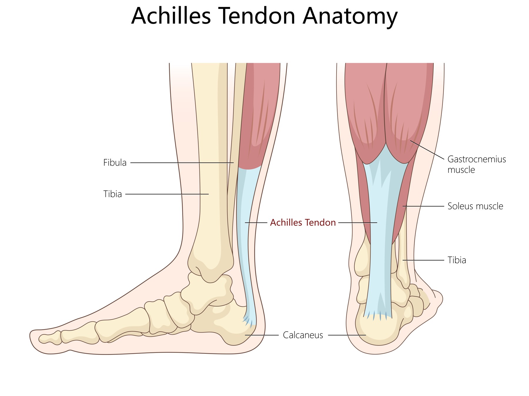
Figure 1. Anatomy of the Achiles tendon. (Licensed from Adobe Stock)
How do Achilles tendon tears happen?
Most Achilles tears are sudden, sports-related injuries. The typical mechanism is a forceful eccentric load, meaning the calf muscle contracts hard while the ankle is simultaneously being pushed up into dorsiflexion. Common scenarios include pushing off to sprint or jump, changing direction quickly, landing awkwardly, or stepping unexpectedly into a hole or off a curb.2,5
A detailed video review of all 77 in-game Achilles ruptures in the National Football League from 2018 to 2024 shows how consistent the pattern is. Sixty-four percent were noncontact injuries and another 35% involved only indirect contact, and they occurred at relatively low running speeds. Three scenarios accounted for 97% of the injuries: change of direction (40%), overload (30%), and rocking back onto the leg (27%). In most cases the injured leg was extended behind the body, with the ankle dorsiflexed and the hip and knee extended.6 In other words, these tears are usually caused by how the leg is loaded, not by being hit.
Most patients describe a sudden snap or pop and often feel as though they were kicked or struck in the back of the ankle, even when no one is near them.2
It is also important to understand that most Achilles tendons that rupture were not entirely normal beforehand. In a controlled study of 891 patients, degenerative changes were present in the great majority of spontaneously ruptured tendons, and roughly one-third of patients report warning symptoms such as calf or heel pain before the injury.2
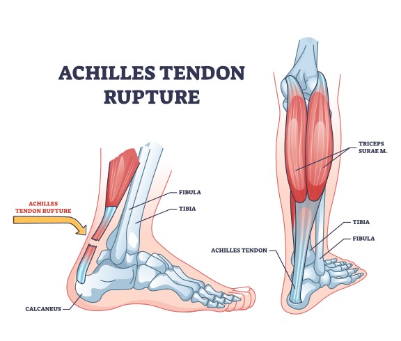
Figure 2. Achilles tendon rupture picture. (Licensed from Adobe Stock)
What are the symptoms of an Achilles Tendon tear?
The most common symptoms are:
- A sudden snap or pop at the back of the ankle, often with the sensation of being kicked or hit2
- Immediate pain at the back of the heel or lower calf, worse with any attempt to push off2
- Swelling and bruising along the back of the ankle over the first day or two
- A palpable gap or divot in the tendon, usually a few centimeters above the heel bone2
- Weakness pushing off, difficulty walking normally, and inability to perform a single-leg heel raise
- An ankle that rests in a less pointed (more dorsiflexed) position than the uninjured side9
One point deserves emphasis. Many patients can still walk after a complete tear, and some can even weakly point the foot down. Other muscles in the leg, including the posterior tibialis, the toe flexors, and the peroneals, can plantar flex the ankle even when the Achilles is completely torn, and injuries to those tendons can produce overlapping symptoms.2,10
With a chronic or neglected tear the pain often improves, but weakness, a limp, difficulty on stairs and hills, and an inability to rise onto the toes persist.11,12
What are common causes of Achilles tendon tears?
The Achilles is the most commonly ruptured tendon in the body.2 In a national analysis of sports-related tendon ruptures presenting to United States emergency departments between 2001 and 2020, Achilles ruptures made up 55.9% of all cases, and basketball was the single most common activity, accounting for 36.6%.13 The reported annual incidence of Achilles rupture ranges from roughly 18 to 37 per 100,000 people, and the number of these injuries has been rising.2,14
Men are affected far more often than women. The injury rate in men was 7.7 times that in women in the emergency department data, and the mean age at injury was 37.7 years.13 Historically the reported mean age has ranged from about 37 to 43.5 years.2
Factors that increase risk include:
- Age-related tendon degeneration and a declining blood supply to the tendon2
- Male sex2,16
- Corticosteroid use, whether oral or injected, and fluoroquinolone antibiotics2
- A sudden change in training, such as a new sport, increased intensity, an unfamiliar surface, or improper footwear2
- High-intensity plyometric or explosive activity2
- Metabolic conditions. In a multicenter study of patients who sustained an Achilles rupture, diabetes, high cholesterol, thyroid disease, and obesity were common.17
- Preexisting Achilles tendinopathy. On MRI, tears located closer to the calf muscle were significantly more likely to occur in tendons showing preexisting tendinopathy.18
- Distance running and other repetitive loading sports19
Once you have torn one Achilles, there is a reported 6% to 26% chance of eventually tearing the other one.2
Tears are also described by location: at the junction of muscle and tendon (myotendinous), in the midsubstance, or at or near the insertion onto the heel bone. A recent MRI-based classification of 428 ruptures found that tear location varies widely and can be measured reliably, that proximal tears were associated with preexisting tendinopathy, and that midsubstance tears were associated with older age and a thicker tendon.18 Insertional tears behave differently from midsubstance tears and are more often associated with degenerative change and calcification.20 Location matters because it influences whether surgery is recommended and which technique is used.21,22
How are Achilles tendon tears diagnosed?
Diagnosis begins with the history and the physical examination, and in most cases the examination alone is enough to make the diagnosis.
On examination I look for:
- A palpable gap in the tendon2
- A positive Thompson test. With you lying face down and the knee bent, I squeeze the calf. Normally this makes the foot point downward. If the tendon is torn, the foot does not move. This test is positive in 96% to 100% of acute ruptures, but in only about 80% of chronic tears.2
- The resting position of the ankle compared with the other side, sometimes measured as the Achilles tendon resting angle. This is useful both at diagnosis and for following tendon length during recovery.9
- Strength testing and, when appropriate, a single-leg heel raise
There are real pitfalls. A large hematoma can fill in the gap and mask it, the other flexor tendons can still point the foot down, and the Thompson test can be falsely negative if the accessory flexors are inadvertently squeezed.
Imaging is used selectively. X-rays are obtained to rule out an avulsion fracture off the heel bone or calcification within the tendon, and to look at the fat pad in front of the tendon (Kager triangle).2 Ultrasound is inexpensive, quick, and allows dynamic assessment.2 MRI gives the most detail: it shows the exact location of the tear, the amount of retraction and the size of the gap, the quality of the remaining tendon, and any partial tearing.2 In one MRI series the average gap measured 30 mm.18 An MRI can be ordered when the diagnosis is unclear.
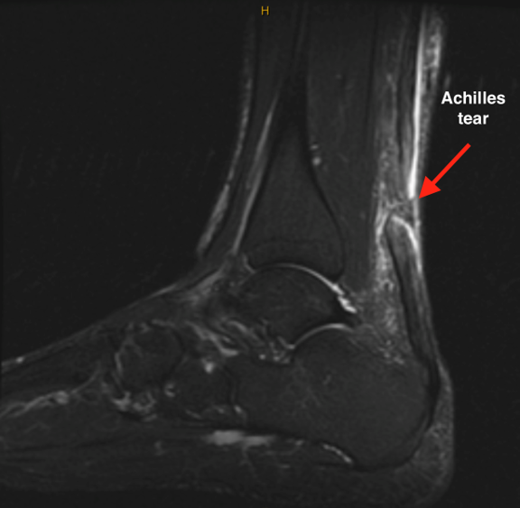
Figure 3. MRI of the ankle demonstrating a complete Achilles tendon rupture.
How are Achilles tendon tears treated?
Treatment is always individually tailored. Considerations include your age, your activity level and the type of activity you want to return to, your occupation, how long ago the injury happened, where the tendon tore, the size of the gap between the tendon ends, the condition of your skin and circulation, other medical conditions such as diabetes or nicotine use, and your goals.1,24
For an acute midsubstance rupture, both nonoperative treatment and surgical repair are reasonable options when either is combined with a modern early functional rehabilitation program.25-27 The trade-off is consistent across the literature: surgery lowers the risk of the tendon tearing again, while nonoperative care avoids the risks that come with an incision. Randomized evidence in this area is also statistically fragile, meaning that in several trials changing the outcome of only a few patients would reverse the conclusion, so the right answer for an individual patient depends heavily on that patient's priorities.28,29 This is one of the most heavily studied topics in foot and ankle surgery, and the literature continues to grow and shift.30,31
Whichever path is chosen, the goal is the same: restore the normal length and tension of the tendon, protect the healing tissue early, and then load it progressively. Tendon length is the outcome that tracks most closely with your final calf strength.32
Nonoperative treatment
Nonoperative treatment means allowing the tendon ends to heal without an incision, using a boot or brace that first holds the ankle pointed downward and then gradually brings it back to neutral. A typical protocol begins with the ankle immobilized in plantar flexion, with heel wedges removed on a set schedule over roughly 8 weeks, and with weightbearing allowed early.2,33
Functional bracing has largely replaced rigid casting. Compared with strict cast immobilization, functional bracing produces better range of motion, an earlier return to preinjury activity, and greater comfort.2 Early weightbearing in a functional orthosis has been reported to give excellent functional outcomes with very low re-rupture rates.33
Modern meta-analyses still show a re-rupture advantage for surgery, but the size of that advantage depends on the rehabilitation protocol used:
- A 2025 meta-analysis of 10 studies and 1327 patients found the re-rupture risk was significantly lower with surgery (relative risk 0.28, 95% CI 0.15-0.50), accompanied by a higher risk of other complications (relative risk 2.39, 95% CI 1.57-3.63).34
- A review of 34 overlapping meta-analyses reported re-rupture rates of 2.3% to 5% with surgery versus 3.9% to 13% with conservative treatment, while conservative treatment had fewer complications overall, in particular fewer infections and fewer sural nerve injuries.25
- A network meta-analysis of 41 studies and 5566 patients reported a re-rupture rate of about 12% with nonsurgical treatment compared with 2% to 4% with surgical treatment.35
- When early range-of-motion protocols are used in both groups, several analyses have found no significant difference in re-rupture.2,26
Function, as opposed to re-rupture, is more similar between the two approaches. In a large Norwegian randomized trial, nonoperative treatment, open repair, and minimally invasive repair did not differ in patient-reported health status at 12 months, although re-rupture within 12 months was more frequent with nonoperative treatment (6.2%) than with open repair (0.6%) or minimally invasive surgery (0.6%).27 A single-institution study using PROMIS scores found that operative and nonoperative groups equalized over time, with more wound-related problems in the operative group.36 In young adults, a midterm study found similar PROMIS scores, blood clot rates, and re-rupture rates between the two treatments.37
Nonoperative treatment does allow the tendon to heal slightly longer in some patients. Using the Achilles tendon resting angle, one study found no clinically relevant difference in tendon elongation between operative and nonoperative treatment at 1 year.9 An MRI study of nonoperatively treated ruptures did document measurable lengthening of the tendon and related it to clinical results.38
Age by itself is not a reason to avoid surgery. A meta-analysis focused on older populations found comparable re-rupture rates between operative and nonoperative management,39 but a prospective study of patients aged 50 and older found significantly better Achilles Tendon Total Rupture Scores at 12 months after percutaneous repair (89.9) than after nonoperative care (74.3), with the largest differences in running, jumping, and heavy physical work.40
Tears at the junction of the muscle and tendon usually do well without surgery. In a systematic review of myotendinous ruptures, all patients were able to perform a single heel raise and returned to work or sport with nonoperative treatment.22 Chronic, neglected ruptures can improve with nonoperative care, but results are generally inferior to surgical reconstruction in active patients.?11,12
Surgical treatment
Surgery is most often chosen by active patients who want the lowest re-rupture risk and the most predictable restoration of tendon length and push-off strength, for larger gaps between the tendon ends, for tears that were not diagnosed acutely, for insertional or avulsion-type tears, and for repeat ruptures.1,24
Minimally invasive techniques were developed specifically to reduce wound problems, and the evidence supports that goal. In a meta-analysis of eight randomized trials, minimally invasive surgery reduced overall complications (relative risk 0.21) and wound infection (relative risk 0.15) compared with open repair, and patients were more likely to report good or excellent subjective results (relative risk 1.18). There was no difference in re-rupture, sural nerve injury, return to preinjury activity level, time to return to work, or ankle range of motion.41 A separate meta-analysis of randomized trials reached similar conclusions.45 In a large series of acute midsubstance repairs the overall complication rate was 15.7% with a major complication rate of 3%, and complications were significantly more common after suture anchor fixation (26.3%) than after percutaneous (14.3%) or open (14.4%) repair.46
Laboratory work supports the strength of these constructs. Cadaveric and finite element testing found the PARS construct comparable to a Krackow repair,51 and biomechanical comparison of nonlocked minimally invasive with locked open repairs helps define how much early loading a repair can tolerate.52 Suture material appears to matter less than technique: absorbable and nonabsorbable sutures showed similar rates of re-rupture, infection, and foreign-body reaction.53 Adding augmentation to a standard repair has not been shown to improve long-term outcome. At a mean of 14 years there was no meaningful difference in outcome score or strength between augmented and nonaugmented repairs.54 When fixation to bone is required, transosseous fixation gives results similar to suture anchors at lower material cost.55
How is an Achilles repair performed?
I most often repair an acute midsubstance Achilles rupture using a mini-open technique with the Arthrex Percutaneous Achilles Repair System (PARS). The goal is to place a strong, locking suture construct into both tendon ends through a small incision. This restores tendon length and tension while leaving the skin envelope and the paratenon as undisturbed as possible, which is the main reason minimally invasive repair has fewer wound complications than open repair.41,45
The steps are as follows:
- The procedure is performed under a general anesthetic, usually with a regional nerve block for postoperative pain control.
- You are positioned face down. The opposite leg is often prepped or positioned so that the resting tension of your normal ankle can be used as a reference when setting the tension of the repair.2
- An incision of roughly 2 to 3 cm is made directly over the rupture site.
- The paratenon, the vascular sleeve around the tendon, is opened carefully and preserved so it can be repaired at the end. The torn tendon ends are then identified.
- The PARS jig is inserted through the incision, deep to the paratenon and alongside the proximal (upper) tendon stump. Needles are passed through the arms of the jig, placing a series of locking and non-locking sutures into the tendon in a whipstitch pattern. The jig is removed, drawing the sutures with it.
- The same is done for the distal (lower) stump. When the distal stump is short, for example with a tear close to the heel bone, the repair can instead be anchored into the calcaneus with a knotless SpeedBridge construct.47
- The ankle is held in plantar flexion, the sutures are tensioned to bring the tendon ends back together, and the repair is tied. Tension is compared with the resting position of the opposite ankle.
- The paratenon is repaired, the skin is closed, and a splint is applied with the ankle in plantar flexion.
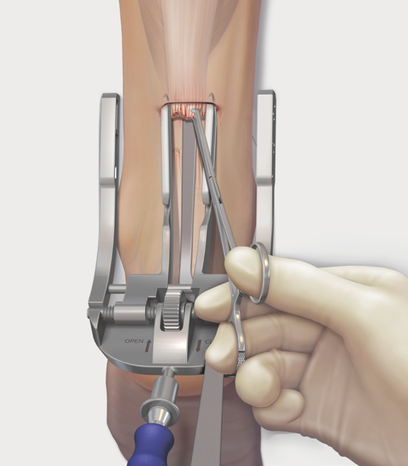
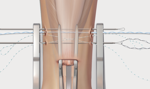
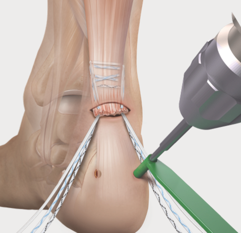
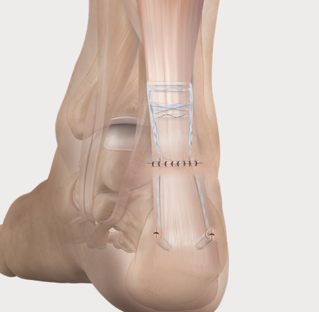
Figure 4. Steps of the Achilles repair procedure. (Credit: Arthrex.com)
What are the outcomes of Achilles tendon repair surgery?
Outcomes after Achilles repair are good to excellent for the large majority of patients.
In the largest comparison of the two minimally invasive systems I use, average scores at a mean of 30 months after surgery were: for PARS, a PROMIS Physical Function score of 58.8, a PROMIS Pain Interference score of 44.2, and an Achilles Tendon Total Rupture Score of 86.0; and for the midsubstance SpeedBridge, 55.3, 44.0, and 82.5 respectively.47 For reference, the PROMIS population mean is 50, so in both groups physical function was above the population average and pain interference was below it.48 Other series report rupture scores in the low 90s after minimally invasive repair, with return-to-sport rates of 91.5% to 92.7%.42
Open and minimally invasive repair give similar patient-reported outcomes.50 Percutaneous knotless repair and open repair give comparable functional outcomes and revision rates.43 Operative and nonoperative treatment converge over time on patient-reported measures, though operative patients may reach population-average physical function sooner.27,36
It is equally important to be honest about what does not fully return. Even after a technically excellent repair, measurable deficits are common:
- At a mean of 14 years after repair, the injured Achilles averaged 12 mm (6%) longer than the uninjured side, soleus and gastrocnemius muscle volumes were 11% to 13% lower, and plantar flexion strength was 12% to 18% lower. The side-to-side difference in tendon length correlated substantially with the strength deficit.32
- Calf muscle volume, strength, and muscle quality remain measurably reduced at 1 year after repair.59
- Heel-rise height and repetitions recover, but patients often use compensatory movement strategies to achieve them.60
- Recovery patterns differ depending on whether the dominant or non-dominant leg was injured.61
- The repaired tendon continues to elongate over the first 6 months regardless of whether weightbearing is started early or late,62 and tendon lengthening changes the mechanics of the foot and reduces push-off efficiency.63
This is precisely why rehabilitation, and calf strengthening in particular, continues well past the point at which the tendon itself has healed.
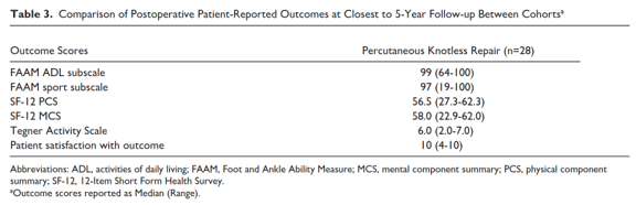
Figure 5. Example of outcome scores. Table from: Stake IK, Matheny LM, Comfort SM, Dornan GJ, Haytmanek CT, Clanton TO. Outcomes Following Repair of Achilles Midsubstance Tears: Percutaneous Knotless Repair vs Open Repair. Foot & Ankle International. 2023;44(6):499-507. doi:10.1177/10711007231160998
When can I expect to return to work and sports after Achilles tendon repair surgery?
Return to work depends far more on what your job requires than on the calendar.
- If your work is sedentary and you can elevate the leg, you may be able to return within the first one to two weeks, once you are off narcotic pain medication and comfortable getting around in the splint with crutches.
- If your job requires standing or walking, return is usually in the 6- to 12-week range as weightbearing progresses and you come out of the boot. In one published protocol, patients were allowed to return to work without restriction between 6 and 12 weeks once their pain and function had improved.22
- Heavy labor, or work on ladders, uneven ground, or at heights, typically requires longer, and light duty is very helpful when it is available.
For sport, the expectation is months rather than weeks.
- In a large minimally invasive series, the average time to return to preinjury level of activity was approximately 9.3 months.48
- A meta-analysis found that return-to-play rates are high overall.66
What are the complications of Achilles tendon repair surgery?
Wound problems and infection
Wound healing complications are the most characteristic risk of Achilles surgery, because the skin over the tendon is thin and its blood supply is limited. Minimally invasive repair substantially reduces this risk; in a meta-analysis of randomized trials the risk ratio for wound infection was 0.15 in favor of minimally invasive surgery compared with open repair.41 In an institutional review of 1148 Achilles operations, 2.0% developed a postoperative wound infection requiring surgical treatment, and among 604 acute repairs specifically the rate was 1.32%.75 Infection can present with increasing pain, redness, warmth, swelling, drainage, and sometimes fever or chills; notify the office for any of these. Management may involve antibiotics, surgical irrigation and debridement, and occasionally staged reconstruction or soft tissue coverage. With early, aggressive treatment most patients recover well, although patients who develop an infection had modestly worse physical function and pain scores at follow-up.75,76 Nicotine dependence significantly increases these risks: in a matched analysis of 2117 nicotine-dependent patients, the risk of wound disruption was increased 1.55-fold and the risk of postoperative infection 1.64-fold within 90 days.77
Re-rupture
Re-rupture after surgical repair occurs in roughly 2% to 5% of patients, compared with roughly 4% to 13% after nonoperative treatment.25,34,35 Pooled across 43 randomized trials, the postoperative re-rupture rate was 3.15%.78 Most re-ruptures occur within the first few months; the median time to re-rupture was 98.5 days in one large series. Risk factors identified at the time of the original injury include male sex, age under 45, and treatment with traditional cast immobilization rather than functional rehabilitation.16 Re-ruptures that occur early after repair can sometimes be treated successfully without surgery, but many require revision surgery.79
Sural nerve injury
The sural nerve supplies sensation to the outer border of the foot and ankle and crosses near the tendon. Injury or irritation is reported in approximately 1% to 2% of minimally invasive repairs, at rates similar to open repair.46,49,50 It usually presents as numbness or tingling along the outside of the foot rather than as weakness, and most cases improve with observation. Persistent painful symptoms occasionally require further treatment.
Blood clots (deep vein thrombosis and pulmonary embolism)
Achilles injuries carry a meaningfully higher risk of blood clots than most orthopaedic problems, because the leg is immobilized in a boot and the normal ankle pump is temporarily lost. In a series of 984 consecutive patients with an acute rupture, most treated nonoperatively, the incidence of symptomatic venous thromboembolism within 90 days was 3.6% (deep vein thrombosis 2.1%, pulmonary embolism 1.9%), and the median time to the event was 24 days. Independent risk factors were age 50 or older and a personal or family history of clot or clotting disorder.80 Early weightbearing appears to reduce the rate of symptomatic events.82 Symptoms include increasing calf or thigh pain and swelling; chest pain or shortness of breath may indicate a pulmonary embolism and is a medical emergency. Prevention includes early motion and weightbearing when permitted, and blood-thinning medication in selected patients.
Tendon lengthening and persistent calf weakness
Even a well-performed repair heals with some lengthening. The ruptured tendon continues to elongate for about 6 months after repair regardless of when weightbearing begins,62 and delaying loading does not eliminate it.83 Lengthening is directly associated with residual weakness32 and alters the mechanics of the foot during push-off.63 In practical terms this can mean persistent difficulty with single-leg heel raises, hills, stairs, and sprinting, and a calf that remains visibly smaller than the other side.59,61
Heel pain and implant-related pain
Constructs that place anchors or knots at the heel bone can cause localized pain there. Suture anchor fixation was associated with clinically significant heel pain in 10.5% of patients, significantly more than percutaneous or open techniques,46 and the midsubstance SpeedBridge showed a higher rate of heel pain at final follow-up than PARS.49 Pain at the heel was also the main driver of the higher complication rate in the PARS and knotless group in another comparative study.42 Most cases improve with time, activity modification, and shoe wear changes. Occasionally implant removal is considered once the tendon has fully healed.
Other complications
Other reported complications include ankle stiffness, scar sensitivity and adhesion of the skin to the tendon, delayed wound healing, prominent or painful suture knots, complex regional pain syndrome, and the general risks of anesthesia. Patients with a diagnosed mental health disorder had higher rates of postoperative infection, postoperative pain, and emergency department visits after Achilles repair, and at longer follow-up higher rates of ankle arthritis and gait abnormality.84
This is not an exhaustive list of all complications. Other complications, although uncommon, can still occur.
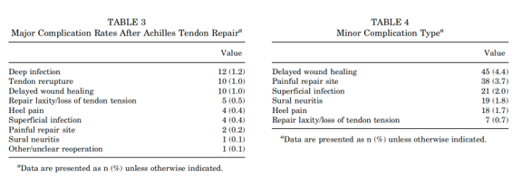
Figure 6. Tables of complications taken from Averkamp BJ, Rees AB, Kalbac T, et al. Comparison of Postoperative Complications by Surgical Technique After Acute Midsubstance Achilles Tendon Repair. The American Journal of Sports Medicine. 2025;53(12):2898-2905. doi:10.1177/03635465251365520
When can I drive after my Achilles tendon surgery?
There is no definitive test that tells us when a specific patient is safe to return to driving. A few principles apply to everyone:
- You should not drive while taking narcotic or other sedating medication.
- You should not drive in a boot or splint. You need to be able to move your foot freely between the pedals and apply full braking force.
- You need to be able to get in and out of the vehicle and control it in an emergency, not simply in ordinary traffic.
The research in this area uses brake reaction time measured on a driving simulator and compared with healthy controls. For a right-sided Achilles repair:
- In a study of 50 patients after open right Achilles repair, brake reaction time returned to safe values in most patients by 6 weeks, and in the remainder by 7 weeks. All patients with a safe brake reaction time at 6 weeks reported feeling ready to drive.95
- A second prospective study of 60 patients reached the same 6- to 7-week conclusion.95
- A survey of 30 patients after open Achilles repair reported an average return to driving at 7 weeks.95
- A review of the overall literature recommends waiting at least 6 to 7 weeks after a right Achilles tendon repair.95
What does rehabilitation look like after an Achilles tear?
Rehabilitation after an Achilles tear is a staged process that lasts roughly a year, and the most important decisions are made in the first 12 weeks.65 The modern approach is early functional rehabilitation: protected weightbearing begun within the first week and controlled exercise begun in the second week, rather than weeks of rigid casting.14 This approach speeds functional recovery without increasing re-rupture, provided motion is progressed in a controlled way.78,96 The protocol below reflects both the published expert consensus and the accelerated functional protocol used in the landmark multicenter randomized trial.65,97
Weeks 0 to 2
Splint or boot with the ankle held in plantar flexion, and non-weightbearing with crutches. Elevate the leg to control swelling. Hip and knee exercises that do not involve the ankle can begin, along with non-weightbearing conditioning such as one-legged cycling.97
Weeks 2 to 4
Transition to a walking boot with heel wedges, and progress to protected weightbearing with crutches. Active plantar flexion and dorsiflexion to neutral only, with no stretching past neutral, plus gentle inversion and eversion below neutral.65,97 Modalities for swelling and incision or scar mobilization are added as the wound allows.97 A stationary bike with the boot on and the heel on the pedal is typically started at an average of 2.4 weeks.65 Formal physical therapy begins in this window.
Weeks 4 to 6
Weightbearing as tolerated in the boot, with the ankle still held in some plantar flexion.65,97 Continue controlled range of motion to neutral, swelling control, and conditioning that does not stress the repair.
Weeks 6 to 8
Heel wedges are removed progressively. Two-legged concentric heel raises typically begin around 6 weeks; the consensus panel's average was 6.4 weeks.65 Graduated resistance exercise, balance and proprioception work, and gait retraining begin, along with weightbearing cardiovascular exercise as tolerated.97
Weeks 8 to 12
Wean out of the boot into a regular shoe with a one-eighth to one-quarter inch heel lift.65 Wean off crutches or a cane. Continue to progress range of motion, strength, and balance.
After 12 weeks
Stretching the ankle past neutral and eccentric (lengthening) calf exercise should not begin before 12 weeks.65 From this point rehabilitation focuses on rebuilding strength, power, and endurance: progressive single-leg heel raises, resistance training, dynamic weightbearing exercise, then plyometric and sport-specific training.97,98
Around 6 months
Return to sport, guided by objective criteria rather than the calendar. Full tendon healing and strength recovery often take 12 months or longer.32,65
Physical therapy should begin within the first 2 to 4 weeks after surgery. Please bring this guide and your operative protocol to your first therapy visit.
Post operative Instructions
- Following your stay in the recovery room and when your vital signs are stable you will be discharged to your escort.
- Remember, it is normal to feel a little dizzy or drowsy for several hours after surgery. This is due to the action the medicine used during surgery.
- If you do not have a post operative appointment scheduled, please call the office as soon as possible to schedule this appointment.
- Take your pain medicine as directed. Begin the pain medicine before you start getting uncomfortable, as the nerve block will wear off. If you wait to take your pain medication until the pain is severe, you will have more difficulty in controlling the pain.
- If you are taking narcotic pain medication you may need a stool softener to prevent constipation. Over-the-counter medication such as Docusate or Milk of Magnesia is recommended.
- Notify the office of any fever, chills, or temperature > 100.5.
- Notify the office of any wound drainage.
Post-Surgery Diet
Resume your diet as tolerated and include vegetables, fruits, and proteins (such as meats, fish, chicken, nuts, and eggs) to promote healing. Also, remember to have adequate fluid intake. It is common after surgery to lack an appetite. This may be the result of anesthesia and the medications. Proper nutrition is needed for healing. During the healing process, the body needs increased amounts of calories and protein. Eat a variety of foods to get all the calories, proteins, vitamins, and minerals you need. If you have been told to follow a specific diet, please follow it.
What to expect after surgery?
You will have a splint that covers the wound(s) on your ankle. You may have some numbness or tingling of foot and ankle due to the nerve block used to help control post-operative pain.
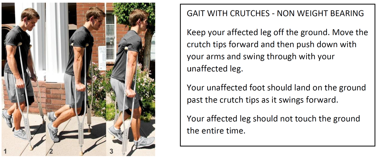
While in the splint work on bending and extending your toes which can help reduce swelling. It is ok to begin to mobilize and walk with the crutches as your pain allows. However, avoid going long periods of time without elevating the leg as this will cause increased pain and swelling.
The splint will be continued for ~2 weeks from the date of the surgery. If the splint begins to feel too tight as a result of swelling it is ok to split the white cast padding underneath the ace bandage and loosen the ace bandage, but do not remove the splint (fiberglass or plaster) material. At the time of your first postoperative appointment you will likely be switched into a CAM boot with wedges.
Dressings & Incisions
- The first two days after surgery you can expect a small amount of red-tinged drainage on your dressings, this is normal.
- Please keep the dressing clean and dry; if you are going to shower/bathe, you must protect the splint from getting wet.
- Do not remove any of the dressings.
- Swelling and bruising in the toes is considered normal.
How do I keep my pain under control?
- It is common to have post-operative pain. You may wrap a bag of frozen peas or crushed ice in a thin towel and place on your dressing for 20 minutes of every hour while you are awake.
- Elevating your ankle above heart level on several pillows will also help with post-operative pain.
- You should take the prescribed medication only as needed and as prescribed.
- If you are taking narcotic pain medication you may need a stool softener to prevent constipation. Over-the-counter medication such as Docusate or Milk of Magnesia is recommended.
- If you had a nerve block at the time of surgery, it usually wears off 12-24 hrs post-operatively. It is normal to have some numbness in the foot the first few days after surgery as a result. The first night after surgery take pain medication before going to bed as the nerve block will often wear off during the night.
Activity
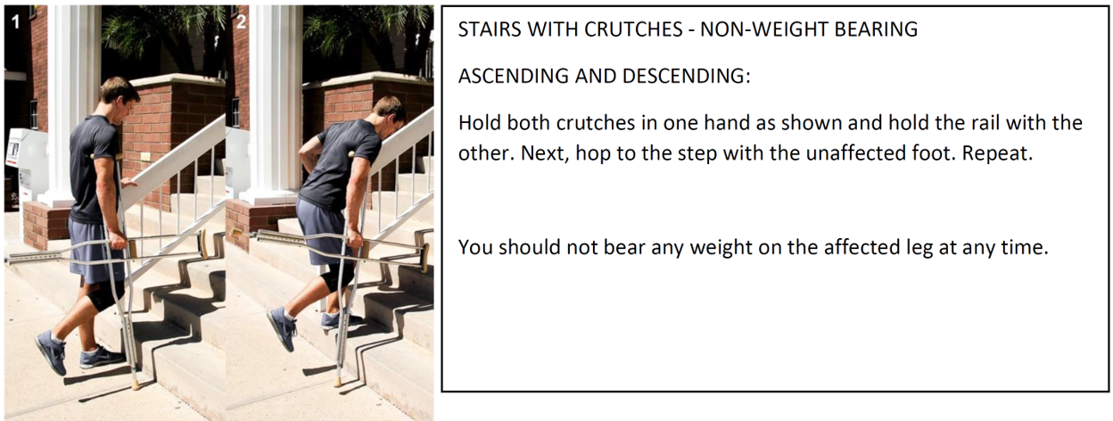
- You may NOT bear weight with your affected leg until cleared by your physician.
- You should move (straighten and bend) your toes at least 10 times per day within your comfort to decrease swelling and prevent stiffness.
References
- Strasser N. Achilles tendon injuries. In: Coughlin and Mann's Surgery of the Foot and Ankle. Elsevier; chap 37:1543-1585. Accessed July 24, 2026. https://www.clinicalkey.com
- Uquillas CA, Guss MS, Ryan DJ, Jazrawi LM, Strauss EJ. Everything Achilles: knowledge update and current concepts in management. J Bone Joint Surg Am. 2015;97(14):1187-1195. doi:10.2106/JBJS.O.00002
- Kim J, Sacks J, El Masry S, Palma J, Ellis SJ, Deland JT. MRI analysis of axial plane rotation and width of the Achilles tendon: implications for minimally invasive Achilles tendon repair. Foot Ankle Orthop. 2024;9(4). doi:10.1177/2473011424S00371
- Mahan J, Damodar D, Trapana E, et al. Achilles tendon complex: the anatomy of its insertional footprint on the calcaneus and clinical implications. J Orthop. 2020;17:221-227. doi:10.1016/j.jor.2019.06.008
- Tarantino D, Palermi S, Sirico F, Corrado B. Achilles tendon rupture: mechanisms of injury, principles of rehabilitation and return to play. J Funct Morphol Kinesiol. 2020;5(4):95. doi:10.3390/jfmk5040095
- Teater RH, Gepner B, O'Cain CM, et al. Video analysis of Achilles tendon ruptures in the National Football League: situational patterns, injury mechanisms, and paths for injury prevention. Am J Sports Med. 2026;54(9):2220-2231. doi:10.1177/03635465261444016
- Cramer A, Hojfeldt G, Schjerling P, et al. Achilles tendon tissue turnover before and immediately after an acute rupture. Am J Sports Med. 2023;51(9):2396-2403. doi:10.1177/03635465231177890
- Nilsson N, Alim MA, Dietrich-Zagonel F, et al. The delayed presentation of Achilles tendon ruptures is associated with marked alterations in the gene expression of COL1A1, MMPs, TIMPs, and IL-6. Am J Sports Med. 2024;52(1):164-173. doi:10.1177/03635465231212669
- Cramer A, Rahdi E, Hansen MS, Sandholdt H, Holmich P, Barfod KW. No clinically relevant difference between operative and non-operative treatment in tendon elongation measured with the Achilles tendon resting angle (ATRA) 1 year after acute Achilles tendon rupture. Knee Surg Sports Traumatol Arthrosc. 2021;29(5):1617-1626. doi:10.1007/s00167-020-06391-w
- Irwin TA. Tendon injuries of the foot and ankle. In: DeLee, Drez, & Miller's Orthopaedic Sports Medicine. Elsevier; chap 118:1462-1483.e5. Accessed July 24, 2026. https://www.clinicalkey.com
- Azam MT, Butler JJ, Weiss MB, et al. Surgical management of chronic Achilles tendon ruptures: a systematic review and proposed treatment algorithm. Foot Ankle Orthop. 2023;8(4). doi:10.1177/24730114231200491
- Arshad Z, Bhatia M. Non-operative management of chronic Achilles tendon ruptures: short term outcomes. Foot Ankle Orthop. 2022;7(1). doi:10.1177/2473011421S00090
- Lyons JG, Berkay FB, Minhas A. Epidemiology of sports-related tendon ruptures presenting to emergency departments in the United States. Am J Sports Med. 2024;52(13):3396-3403. doi:10.1177/03635465241284644
- Zellers JA, Christensen M, Kjaer IL, Rathleff MS, Gravare Silbernagel K. Defining components of early functional rehabilitation for acute Achilles tendon rupture: a systematic review. Orthop J Sports Med. 2019;7(11):2325967119884071. doi:10.1177/2325967119884071
- Ho G, Tantigate D, Kirschenbaum J, Greisberg JK, Vosseller JT. Increasing age in Achilles rupture patients over time. Injury. 2017;48(7):1701-1709. doi:10.1016/j.injury.2017.04.007
- Maempel JF, White TO, Mackenzie SP, McCann C, Clement ND. The epidemiology of Achilles tendon re-rupture and associated risk factors: male gender, younger age and traditional immobilising rehabilitation are risk factors. Knee Surg Sports Traumatol Arthrosc. 2022;30(7):2457-2469. doi:10.1007/s00167-021-06824-0
- Oliva F, Marsilio E, Asparago G, et al. Achilles tendon rupture and dysmetabolic diseases: a multicentric, epidemiologic study. J Clin Med. 2022;11(13):3698. doi:10.3390/jcm11133698
- Mueller MM, Estfeller A, Cychosz CC, et al. MRI-based classification of Achilles tendon ruptures: reliability and predictors of tear location. Orthop J Sports Med. Published online 2026. doi:10.1177/23259671261428092
- Jastifer JR. Contemporary review: the foot and ankle in long-distance running. Foot Ankle Orthop. 2022;7(3). doi:10.1177/24730114221125455
- Ramakanth R, Sundararajan SR, Goud BH, Dsouza T, Rajasekaran S. Insertional versus non-insertional tendoachilles tears: a comparative analysis of various predisposing factors and outcome following a repair. Int Orthop. 2022;46(5):1009-1017. doi:10.1007/s00264-022-05337-w
- Giordano J, Partan M, Iturriaga C, Granata J, Katsigiorgis G, Cohn R, Bitterman A. The relationship between patient demographics, tear locations, and operative techniques on the surgical treatment of acute Achilles tendon ruptures. Cureus. 2022;14(8):e28300. doi:10.7759/cureus.28300
- Lameire DL, Ramelli L, Halai M, Wasserstein D, Park SS. Outcomes of operative and nonoperative management of myotendinous Achilles tendon ruptures: a systematic review. BMC Musculoskelet Disord. 2025;26(1):71. doi:10.1186/s12891-025-08286-8
- Arif HA, Devkumar D, Sultan A, Williams KA, Johnson MD. Retrospective analysis of malpractice claims following Achilles tendon rupture. Foot Ankle Orthop. 2025;10(4). doi:10.1177/24730114251397974
- Cohen D, Sandman E, Saran N, Petrisor B, Bhandari M. Evidence-based treatment of Achilles tendon rupture. Can J Surg. 2023;66(4):E356-E358. doi:10.1503/cjs.008321
- Seow D, Islam W, Randall GW, et al. Lower re-rupture rates but higher complication rates following surgical versus conservative treatment of acute Achilles tendon ruptures: a systematic review of overlapping meta-analyses. Knee Surg Sports Traumatol Arthrosc. 2023;31(9):3528-3540. doi:10.1007/s00167-023-07411-1
- Reito A, Mattila V, Karjalainen T. Operative vs nonoperative treatment of Achilles tendon ruptures using early functional rehabilitation: critical analysis of evidence. Foot Ankle Int. 2022;43(7):887-890. doi:10.1177/10711007221083691
- Myhrvold SB, Brouwer EF, Andresen TKM, et al. Nonoperative or surgical treatment of acute Achilles' tendon rupture. N Engl J Med. 2022;386(15):1409-1420. Abstracted in: J Bone Joint Surg Am. 2022;104(22):2035.
- Doyle TR, Davey MS, Hurley ET. The statistical fragility of management options for acute Achilles tendon ruptures: a systematic review of randomized control trial with fragility analysis. J ISAKOS. 2022;7(3):72-81. doi:10.1016/j.jisako.2022.04.003
- Bragg JT, Ruelos VCB, McIntyre JA, et al. Reverse fragility index comparing rates of rerupture after open Achilles tendon repair versus early functional rehabilitation: a systematic review of randomized controlled trials. Am J Sports Med. 2024;52(4):1116-1121. doi:10.1177/03635465231178831
- Markel JF, Anciano V. Achilles tendon rupture - the essentials: a bibliometric study. Foot Ankle Orthop. 2023;8(4). doi:10.1177/2473011423S00225
- Meulenkamp B, Stacey D, Fergusson D, Hutton B, Shorr R, Graham ID. Protocol for treatment of Achilles tendon ruptures: a systematic review with network meta-analysis. Syst Rev. 2018;7(1):247. doi:10.1186/s13643-018-0912-5
- Heikkinen J, Lantto I, Piilonen J, et al. Tendon length, calf muscle atrophy, and strength deficit after acute Achilles tendon rupture: long-term follow-up of patients in a previous study. J Bone Joint Surg Am. 2017;99(18):1509-1515. doi:10.2106/JBJS.16.01491
- Naskar R, Oliver L, Velazquez-Ruta P, Dhinsa B, Southgate C. Functional outcome of early weight bearing for acute Achilles tendon rupture treated conservatively in a weight-bearing orthosis. Foot Ankle Surg. 2022;28(5):595-602. doi:10.1016/j.fas.2021.06.008
- Espana Fernandez de Valderrama S, Garcia Martinez B, Ezquerra Herrando L. Achilles tendon rupture treatment: systematic review and meta-analysis. Foot Ankle Orthop. 2025;10(2). doi:10.1177/24730114251327219
- Pisano A, Boxler M, Gambuti E, et al. Open surgical repair as gold standard for acute Achilles tendon ruptures: systematic review and network meta-analysis. Knee Surg Sports Traumatol Arthrosc. 2025;33(7):2664-2683. doi:10.1002/ksa.12686
- Burger P, Botros M, El-Zein Z, Holleran A, Ketz J, Flemister AS, Ciufo D. PROMIS outcomes after operative vs nonoperative treatment of Achilles rupture. Foot Ankle Int. 2025;46(7):715-722. doi:10.1177/10711007251336756
- Ge L, Saunders N, Betts D, Holmes JR, Walton DM, Talusan PG. Midterm outcomes of operatively and nonoperatively managed Achilles tendon ruptures in young adults. Foot Ankle Orthop. 2023;8(3). doi:10.1177/24730114231198849
- Yasuda T, Togei K, Tanaka K, Takashima M, Shima H, Otsuki S. Non-operative treatment for acute Achilles tendon rupture: evaluation of clinical results and tendon lengthening on magnetic resonance imaging. Foot Ankle Orthop. 2026;11(2). doi:10.1177/24730114261436395
- Acevedo D, Garcia JR, Grewal RS, et al. Comparison of rerupture rates after operative and nonoperative management of Achilles tendon rupture in older populations: systematic review and meta-analysis. J Orthop. 2024;52:112-118. doi:10.1016/j.jor.2024.02.034
- Sadiq S, Grocott N, Ibrahim A, et al. Percutaneous repair in patients aged 50 years and older is associated with good outcomes in acute Achilles tendon rupture. Foot Ankle Orthop. 2026;11(1). doi:10.1177/24730114251413239
- Grassi A, Amendola A, Samuelsson K, et al. Minimally invasive versus open repair for acute Achilles tendon rupture: meta-analysis showing reduced complications, with similar outcomes, after minimally invasive surgery. J Bone Joint Surg Am. 2018;100(22):1969-1981. doi:10.2106/JBJS.17.01364
- Goveo MA, Crosa JI, Peralta JI, Colombo C, Vago Anaya F, Loncharich E. Minimally invasive Achilles tendon repair: Dresden vs PARS/knotless techniques in a retrospective comparative study. Foot Ankle Int. 2025;46(9):951-957. doi:10.1177/10711007251341896
- Stake IK, Matheny LM, Comfort SM, Dornan GJ, Haytmanek CT, Clanton TO. Outcomes following repair of Achilles midsubstance tears: percutaneous knotless repair vs open repair. Foot Ankle Int. 2023;44(6):499-507. doi:10.1177/10711007231160998
- Maffulli N, Ziello S, Maisto G, Migliorini F, Oliva F. Local tendon transfers for chronic ruptures of the Achilles tendon: a systematic review. J Clin Med. 2023;12(2):707. doi:10.3390/jcm12020707
- Attia AK, Mahmoud K, d'Hooghe P, Bariteau J, Labib SA, Myerson MS. Outcomes and complications of open versus minimally invasive repair of acute Achilles tendon ruptures: a systematic review and meta-analysis of randomized controlled trials. Am J Sports Med. 2023;51(3):825-836. doi:10.1177/03635465211053619
- Averkamp BJ, Rees AB, Kalbac T, et al. Comparison of postoperative complications by surgical technique after acute midsubstance Achilles tendon repair. Am J Sports Med. 2025;53(12):2898-2905. doi:10.1177/03635465251365520
- Peabody JJ, Hadley SM Jr, Bergman R, et al. Functional outcomes following minimally invasive Achilles rupture repair: a retrospective comparative study of PARS and midsubstance speedbridge techniques. J Orthop Surg Res. 2025;20(1):390. doi:10.1186/s13018-025-05748-6
- Peabody JJ, Hadley SM, Westvold S, Olamigoke F, Chang S, Patel M, Kadakia AR. Patient reported outcomes of minimally invasive Achilles rupture repair. Foot Ankle Orthop. 2024;9(4). doi:10.1177/2473011424S00419
- Peabody JJ, Hadley SM, Westvold S, Devlin S, Chang S, Patel M, Kadakia AR. Complications of minimally invasive Achilles repair using the midsubstance speedbridge and percutaneous Achilles repair system. Foot Ankle Orthop. 2024;9(4). doi:10.1177/2473011424S00216
- Caolo KC, Eble SK, Rider C, et al. Clinical outcomes and complications with open vs minimally invasive Achilles tendon repair. Foot Ankle Orthop. 2021;6(4). doi:10.1177/24730114211060063
- Macaluso B, Hassan CR, Swanson DR, et al. Biomechanical comparison of Krackow repair and percutaneous Achilles repair system for Achilles tendon rupture fixation: a cadaveric and finite element analysis study. Foot Ankle Orthop. 2022;7(1). doi:10.1177/24730114221088502
- Chuckpaiwong B, Glisson RR, Usuelli FG, Madi NS, Easley ME. Biomechanical comparison of nonlocked minimally invasive and locked open Achilles tendon simulated rupture repairs. Foot Ankle Int. 2023;44(9):913-921. doi:10.1177/10711007231178819
- Oesman I, Canintika AF. Absorbable vs nonabsorbable sutures for Achilles tendon repair: a systematic review and meta-analysis. Foot Ankle Orthop. 2023;8(4). doi:10.1177/24730114231201842
- Heikkinen J, Lantto I, Flinkkila T, Ohtonen P, Pajala A, Siira P, Leppilahti J. Augmented compared with nonaugmented surgical repair after total Achilles rupture: results of a prospective randomized trial with thirteen or more years of follow-up. J Bone Joint Surg Am. 2016;98(2):85-92. doi:10.2106/JBJS.O.00496
- Wolf J, Fallat L, Coffey M. Outcomes and material cost comparison of transosseous versus suture anchor fixation of the Achilles tendon: a retrospective study. J Foot Ankle Surg. 2022;61(1):12-16. doi:10.1053/j.jfas.2021.05.012
- Yasuda T, Shima H, Mori K, Kizawa M, Neo M. Direct repair of chronic Achilles tendon ruptures using scar tissue located between the tendon stumps. J Bone Joint Surg Am. 2016;98(14):1168-1175. doi:10.2106/JBJS.15.00865
- Murdock CJ, Ochuba AJ, Xu AL, Snow M, Bronheim R, Vulcano E, Aiyer AA. Operative vs nonoperative management of Achilles tendon rupture: a cost analysis. Foot Ankle Orthop. 2023;8(1). doi:10.1177/24730114231156410
- Wagers K, Ofori-Atta BS, Tucker W, Presson AP, Nixon D. Evaluation of costs associated with acute Achilles tendon repair. Foot Ankle Orthop. 2024;9(1). doi:10.1177/24730114241238215
- Mashimo S, Nozaki T, Amaha K, Tanaka K, Kubota J, Sato H, Kitamura N. Quantitative assessment of calf muscle volume, strength, and quality after Achilles tendon rupture repair: a 1-year prospective follow-up study. Am J Sports Med. 2023;51(14):3781-3789. doi:10.1177/03635465231206391
- Aufwerber S, Gravare Silbernagel K, Ackermann PW, Naili JE. Comparable recovery and compensatory strategies in heel-rise performance after a surgically repaired acute Achilles tendon rupture: an in vivo kinematic analysis comparing early functional mobilization and standard treatment. Am J Sports Med. 2022;50(14):3856-3865. doi:10.1177/03635465221129284
- Xiao X, Kim Y, Lee J. Comparison of dominant and non-dominant muscle function recovery and limb symmetry after Achilles tendon repair: a retrospective study. J Clin Med. 2026;15(2):707. doi:10.3390/jcm15020707
- Eliasson P, Agergaard AS, Couppe C, et al. The ruptured Achilles tendon elongates for 6 months after surgical repair regardless of early or late weightbearing in combination with ankle mobilization: a randomized clinical trial. Am J Sports Med. 2018;46(10):2492-2502. doi:10.1177/0363546518781826
- Malakoutikhah H, Latt LD. The impact of tendon lengthening following Achilles tendon repair on foot kinetics, a finite element analysis. Foot Ankle Orthop. 2023;8(4). doi:10.1177/2473011423S00429
- Brumann M, Baumbach SF, Mutschler W, Polzer H. Accelerated rehabilitation following Achilles tendon repair after acute rupture: development of an evidence-based treatment protocol. Injury. 2014;45(11):1782-1790. doi:10.1016/j.injury.2014.06.022
- Saxena A, Giai Via A, Gravare Silbernagel K, et al. Current consensus for rehabilitation protocols of the surgically repaired acute mid-substance Achilles rupture: a systematic review and recommendations from the “GAIT” study group. J Foot Ankle Surg. 2022;61(4):855-861. doi:10.1053/j.jfas.2021.12.008
- Bak BM, Seow D, Teo YZE, Hasan MY, Pearce CJ. Return to play and functional outcomes following treatment of acute Achilles tendon ruptures: a systematic review and meta-analysis. J Foot Ankle Surg. 2024;63(3):420-429. doi:10.1053/j.jfas.2023.12.008
- Lezak BA, Butler JJ, Phadke R, et al. Outcomes following Achilles tendon ruptures in the National Hockey League: a retrospective sports database study. J Clin Med. 2025;14(15):5471. doi:10.3390/jcm14155471
- Aiyer AA, Hodgens BH, Geller JS, Rizzo MG, Munoz J, Kaplan JR. Performance outcomes after surgical repair of Achilles tendon rupture in the Women's National Basketball Association. Foot Ankle Orthop. 2022;7(1). doi:10.1177/2473011421S00074
- Diniz P, Lacerda D, Mendes B, Pereira H, Ferreira FC, Kerkhoffs GMMJ. Return-to-performance in elite soccer players after Achilles tendon ruptures: a study using a weighted plus/minus metric and matched-control analysis. Knee Surg Sports Traumatol Arthrosc. 2023;31(12):6059-6068. doi:10.1007/s00167-023-07607-5
- Wright CJ, Robinson ABJ, Waldrop NE III, Anderson RB, Clanton TO. Are prospective criteria or objective clinical measures utilized in return to play (RTP) decision making after ankle surgery? A scoping review. Foot Ankle Orthop. 2023;8(1). doi:10.1177/24730114231160996
- Lopes R, Freiha K, Carmont MR, et al. Validation of a composite outcome score for assessing return to sports after Achilles tendon repair. Am J Sports Med. 2025;53(7):1707-1715. doi:10.1177/03635465251333142
- Shitrit E, Valentin E, Baudrier N, et al. The ALR-RSI score can be used to evaluate psychological readiness to return to sport after acute Achilles tendon tear. Knee Surg Sports Traumatol Arthrosc. 2023;31(11):4961-4968. doi:10.1007/s00167-023-07548-z
- Hadley SM Jr, Peabody JJ, Bergman R, et al. Fear of reinjury is associated with worse physical function measured by PROMIS following minimally invasive Achilles rupture repair. Foot Ankle Orthop. 2026;11(3). doi:10.1177/24730114261462481
- Jonsdottir US, Brorsson A, Nilsson Helander K, Tranberg R, Larson MEH. Factors that affect return to sports after an Achilles tendon rupture: a qualitative content analysis. Orthop J Sports Med. 2023;11(1). doi:10.1177/23259671221145199
- Kukadia SK, Cho DJ, Burn CP, Gamarra JA, Rider CM, Henry M, Drakos MC. Management and functional outcomes after postoperative Achilles tendon infection: a retrospective case series. Foot Ankle Int. 2025;46(12):1377-1385. doi:10.1177/10711007251379863
- Bae SH, Lee HS, Seo SG, Kim SW, Gwak HC, Bae SY. Debridement and functional rehabilitation for Achilles tendon infection following tendon repair. J Bone Joint Surg Am. 2016;98(14):1161-1167. doi:10.2106/JBJS.15.01117
- DeShazo S, Pescatore SM, Panchbhavi VK. Nicotine dependence and rates of postoperative complications in Achilles tendon repair. Foot Ankle Int. 2024;45(1):67-72. doi:10.1177/10711007231205293
- Wang R, Huang L, Jiang S, et al. Immediate mobilization after repair of Achilles tendon rupture may increase the incidence of re-rupture: a systematic review and meta-analysis of randomized controlled trials. Int J Surg. 2024;110(6):3888-3899. doi:10.1097/JS9.0000000000001305
- Choi JY, Suh JS, Kim MJ. Conservative treatment outcomes for Achilles tendon re-rupture occurring in the subacute phase after primary repair. Foot Ankle Orthop. 2023;8(4). doi:10.1177/2473011423S00272
- Oliver WM, Mackenzie SA, Lenart L, et al. Age, personal and family history are independently associated with venous thromboembolism following acute Achilles tendon rupture. Injury. 2022;53(2):762-770. doi:10.1016/j.injury.2021.10.009
- Aufwerber S, Svedman S, Gravare Silbernagel K, Ackermann PW. Long-term patient outcome is affected by deep venous thrombosis after Achilles tendon rupture repair. Knee Surg Sports Traumatol Arthrosc. 2024;32(9):2184-2193. doi:10.1002/ksa.12240
- Lee C, Haarer F, Titheradge R, Iliopoulos E. Thromboembolic events during weightbearing vs nonweightbearing accelerated rehabilitation protocols for complete Achilles tendon ruptures. Foot Ankle Orthop. 2023;8(2). doi:10.1177/24730114231178791
- Hoeffner R, Agergaard AS, Svensson RB, et al. Tendon elongation and function after delayed or standard loading of surgically repaired Achilles tendon ruptures: a randomized controlled trial. Am J Sports Med. 2024;52(4):1022-1031. doi:10.1177/03635465241227178
- Kesaria AA, Irfan SM, Nguyen TX, Shi JL, Ruelos VCB, Weiss WM. Impact of diagnosed mental health disorders on postoperative outcomes after Achilles tendon repair. Foot Ankle Orthop. 2026;11(2). doi:10.1177/24730114261457093
- Pretorius J, Habash M, Ghobrial B, Alnajjar R, Ellanti P. Current status and advancements in platelet-rich plasma therapy. Cureus. 2023;15(10):e47176. doi:10.7759/cureus.47176
- Rothrauff BB, Featherall JT, Uppstrom TJ, et al. Platelet-rich plasma in the treatment of musculoskeletal disease in 2025 and beyond. Am J Sports Med. 2026;54(8):2058-2074. doi:10.1177/03635465251395284
- Keene DJ, Alsousou J, Harrison P, et al; PATH-2 Trial Group. Platelet rich plasma injection for acute Achilles tendon rupture: PATH-2 randomised, placebo controlled, superiority trial. BMJ. 2019;367:l6132. Abstracted in: J Bone Joint Surg Am. 2020;102(16):1466.
- Boesen A, Boesen M, Hansen R, Weisskirchner Barfod K, Lenskjold A, Malliaras P, Langberg H. Effect of platelet-rich plasma on nonsurgically treated acute Achilles tendon ruptures: a randomized, double-blinded prospective study. Am J Sports Med. 2020;48(9):2268-2276. Abstracted in: J Bone Joint Surg Am. 2021;103(10):934.
- Boksh K, Elbashir M, Thomas O, Divall P, Mangwani J. Platelet-rich plasma in acute Achilles tendon ruptures: a systematic review and meta-analysis. Foot (Edinb). 2022;53:101923. doi:10.1016/j.foot.2022.101923
- Abdolrazaghi S, Akram N, Boksh K, Elbashir M, Thomas O, Divall P, Mangwani J. Platelet-rich plasma in acute Achilles tendon ruptures: a systematic review and meta-analysis. Foot Ankle Orthop. 2022;7(4). doi:10.1177/2473011421S00549
- Nelson PA, George T, Bowen E, Sheean AJ, Bedi A. An update on orthobiologics: cautious optimism. Am J Sports Med. 2023;51(12):3196-3201. doi:10.1177/03635465231192473
- Bachir RM, Zaia IM, Santos GS, et al. Bone marrow aspirate concentrate improves outcomes in adults with osteochondral dissecans of the talus and Achilles rupture. Arthroscopy. 2023;39(3):881-886. doi:10.1016/j.arthro.2022.11.034
- Baker KC, Baker EA, Fortin PT, Vaupel ZM, Loy BN. Influence of local chemokine delivery and postoperative peripheral blood mobilization on Achilles tendon repair. Foot Ankle Orthop. 2022;7(1). doi:10.1177/2473011421S00103
- Wei K, Deng XH, Mou Y, Wang T, Sun X, Nie M. Comparative efficacy of surgical versus nonsurgical management for acute Achilles tendon rupture in a novel mouse model. J Bone Joint Surg Am. 2026;108(12):929-938. doi:10.2106/JBJS.25.01211
- Frane N, Bandovic I, Hu V, Bitterman A. Return-to-driving recommendations after lower-extremity orthopaedic procedures. JBJS Rev. 2020;8(12):e20.00066. doi:10.2106/JBJS.RVW.20.00066
- Gould HP, Bano JM, Akman JL, Fillar AL. Postoperative rehabilitation following Achilles tendon repair: a systematic review. Sports Med Arthrosc Rev. 2021;29(3):130-145.
- Willits K, Amendola A, Bryant D, Mohtadi NG, Giffin JR, Fowler P, Kean CO, Kirkley A. Operative versus nonoperative treatment of acute Achilles tendon ruptures: a multicenter randomized trial using accelerated functional rehabilitation. J Bone Joint Surg Am. 2010;92(17):2767-2775. doi:10.2106/JBJS.I.01401
- Marrone W, Andrews R, Reynolds A, Vignona P, Patel S, O'Malley M. Rehabilitation and return to sports after Achilles tendon repair. Int J Sports Phys Ther. 2024;19(9):1152-1165. doi:10.26603/001c.122643
- Lee KW, Bae JY, Ho BC, Kim JH, Seo DK. Immediate weightbearing and ankle motion exercise after acute Achilles tendon rupture repair. J Foot Ankle Surg. 2022;61(3):604-608. doi:10.1053/j.jfas.2021.10.021
- Maffulli N, Gougoulias N, Maffulli GD, Oliva F, Migliorini F. Slowed-down rehabilitation following percutaneous repair of Achilles tendon rupture. Foot Ankle Int. 2022;43(2):244-252. doi:10.1177/10711007211038594
- Rendek Z, Bon Beckman L, Schepull T, Danmark I, Aspenberg P, Schilcher J, Eliasson P. Early tensile loading in nonsurgically treated Achilles tendon ruptures leads to a larger tendon callus and a lower elastic modulus: a randomized controlled trial. Am J Sports Med. 2022;50(12):3286-3298. doi:10.1177/03635465221117780
- Roell AE, Timmers TK, van der Ven DJC, van Olden GDJ. Rehabilitation after surgical repair of acute Achilles tendon rupture: functional outcome with a minimum follow-up of 6 months. J Foot Ankle Surg. 2021;60(3):482-488. doi:10.1053/j.jfas.2020.09.003


