
Ankle Fracture Specialist in Georgetown, Cedar Park, Round Rock, TX
Ankle fracture is a condition where there is a break in one or more bones forming the ankle joint. It occurs from excessive rolling and twisting of the ankle often resulting in swelling, severe pain around the ankle and impaired mobility. Arturo Villarreal M.D., Board Certified Orthopaedic Surgeon provides expert diagnosis and individualized non-operative and operative treatments for ankle fractures in Georgetown, Cedar Park, Round Rock, TX. Contact Arturo Villarreal M.D., Board Certified Orthopaedic Surgeon’s team for an appointment today!


What is the ankle?
The ankle is the joint that connects your leg to your foot. It is formed by three bones: the tibia (shin bone), the fibula (the thinner bone along the outside of the leg), and the talus, which sits between them and connects down to the heel bone.1 The bottom end of the tibia forms the medial malleolus, the bump you can feel on the inside of your ankle. The bottom end of the fibula forms the lateral malleolus, the bump on the outside, which extends about a centimeter lower and further back than the medial side. The back edge of the tibia is called the posterior malleolus.1 Together these three “malleoli” wrap around the talus like a socket. This socket is called the ankle mortise, and it works as a modified hinge that lets the foot move up and down.1
The bones alone do not keep the ankle stable. Strong ligaments hold the joint together, and they are just as important as the bone.2 On the inside of the ankle is the deltoid ligament, which has a superficial layer and a deep layer. The superficial layer keeps the heel from rolling outward, and the deep layer keeps the talus from rotating within the mortise.1 On the outside of the ankle are three ligaments: the anterior talofibular ligament, the calcaneofibular ligament, and the posterior talofibular ligament. The anterior talofibular ligament is the weakest of the three and the one most often injured.1
Just above the joint, a group of four ligaments called the syndesmosis binds the tibia and fibula together. The syndesmosis keeps the two leg bones from spreading apart when you put weight through the ankle.1 Because the ankle depends on both bone and ligament, an injury can break bone, tear ligament, or do both. Which structures are damaged is what ultimately determines whether an ankle fracture is stable or unstable, and therefore how it should be treated.2,3
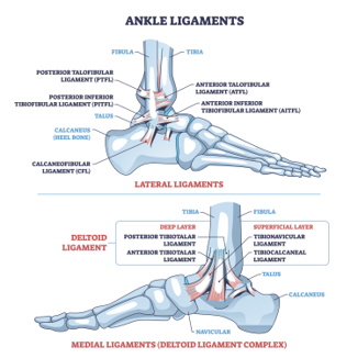
Figure 1. The ligaments of the ankle. The top image shows the lateral (outside) ligaments and the syndesmotic ligaments that hold the tibia and fibula together. The bottom image shows the deltoid ligament complex on the medial (inside) of the ankle, with its deep and superficial layers. (licensed from Adobe Stock)
How is the ankle fractured?
Most ankle fractures happen when the foot is planted on the ground and the body rotates over it. The talus twists inside the mortise and forces the malleoli and ligaments past the point where they can hold, which is why these are often called rotational ankle fractures.1 In the Swedish Fracture Register, which captured more than 57,000 ankle fractures over ten years, a simple fall was the single most common mechanism for every fracture type studied.4
Broken down further, ankle fractures have been reported to result from a fall in about 36% of cases, a sports injury in 35%, an exercise-related injury in 19%, a jump in 5%, and generalized trauma in 4%.1 There is also a seasonal pattern, with more ankle fractures occurring during the winter months, likely reflecting ice and slippery surfaces.4 Newer causes continue to appear as well; electric scooter accidents have become a documented source of foot and ankle injuries.5
What are the symptoms of an ankle fracture?
The most common symptoms are pain, swelling, and bruising around the ankle. Pain is typically worse with any attempt to move the ankle or put weight on it. Tenderness located directly over one of the malleoli, combined with an inability to bear weight, is one of the strongest indicators that a fracture is present and that x-rays should be obtained.2
If the fracture is displaced or the ankle has dislocated, there may be an obvious deformity, with the foot sitting in an abnormal position relative to the leg. This is important to recognize quickly. A markedly deformed ankle stretches the skin over the bone, promotes further swelling, and can interfere with blood flow to the foot, so it should be reduced and splinted urgently.2 Numbness, a pale or dusky foot, or an inability to feel the foot are all reasons to seek care immediately.2
It is also worth knowing what is normal later on. Soft tissue swelling around the ankle frequently persists for months after the injury, well after the bone itself has healed, and this by itself is not a sign that something has gone wrong.2
How common is an ankle fracture?
Ankle fractures are among the most common fractures treated by orthopedic surgeons. They are the third most common type of fracture, affecting both sexes and all age groups,4 and account for roughly 9% of all fractures.1 In Sweden, where fractures are tracked in a national register, the incidence in 2021 was approximately 152 ankle fractures per 100,000 adults per year.4 In a large United States insurance claims analysis, ankle fractures affected 0.14% of the population, and about 23% of those fractures went on to require surgery.6
How are ankle fractures classified?
Several classification systems are used, and your surgeon may use more than one when describing your injury.
The simplest and most widely used is the Danis-Weber classification, which is based on where the fibula is broken relative to the syndesmosis. A Weber A fracture occurs below the syndesmosis, a Weber B fracture occurs at the level of the syndesmosis, and a Weber C fracture occurs above it.1 This matters because the higher the fibula fracture, the more likely the syndesmotic ligaments have also been injured, and the more likely the ankle is unstable. The Danis-Weber system is simpler than the alternatives and has better agreement between different observers.1 In the Swedish Fracture Register, 24% of ankle fractures were type A, 64% were type B, and 12% were type C.4
The Lauge-Hansen classification describes the position of the foot at the time of injury and the direction of the force that caused it. It is useful for describing fracture patterns, but it can only classify about 60% of ankle fractures and agreement between observers is poor.1 The AO/OTA classification builds on the Danis-Weber system and adds descriptions of the medial side of the ankle.1
Separately, fractures are commonly described by how many malleoli are broken. A unimalleolar fracture involves one, a bimalleolar fracture involves two, and a trimalleolar fracture involves all three. In a Finnish national study of more than 83,000 surgically treated ankle fractures, 36% were isolated lateral malleolar fractures, 7% were isolated medial malleolar fractures, and 52% were bimalleolar or trimalleolar.7 A Maisonneuve fracture is a specific pattern in which the fibula breaks high up near the knee and the injury travels down through the syndesmosis to the ankle; this is easy to miss unless the entire leg is imaged.2
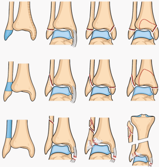
Figure 2. Common ankle fracture patterns arranged by the level of the fibula fracture. The top row shows type A (Weber A) injuries below the syndesmosis, the middle row shows type B (Weber B) injuries at the level of the syndesmosis, and the bottom row shows type C (Weber C) injuries above the syndesmosis, including a high fibula (Maisonneuve) fracture. Within each row, the pattern progresses from an isolated fibula fracture to injuries that also involve the medial malleolus and the posterior malleolus.
How are ankle fractures diagnosed?
During the clinic visit, x-rays will be obtained. The standard series includes three views: an anteroposterior view, a lateral view, and a mortise view. If there is tenderness higher up the leg, full-length views of the fibula are essential to rule out a Maisonneuve injury.2
Your x-rays are used to take several measurements rather than simply to confirm that a bone is broken. These include the medial clear space, which is the gap between the medial malleolus and the talus; the tibiofibular clear space and overlap, which reflect the integrity of the syndesmosis; and the talar tilt.1,8 Widening of the medial clear space beyond about 4 mm suggests the talus has shifted within the mortise and that the ankle is unstable.1
Some fractures look stable on a resting x-ray but are not. To sort this out, additional tests may be used. Stress x-rays, taken either by letting gravity pull the foot outward or by having the examiner rotate the foot, can reveal instability that is otherwise hidden; both methods perform similarly for surgical decision-making.10 A weight-bearing x-ray, in which you are asked to stand and take a few steps, is another simple and reliable way to identify a stable Weber B fracture that can be treated without surgery.11
Advanced imaging is used selectively. A CT scan gives a cross-sectional view of the joint and is particularly useful for sizing a posterior malleolus fracture, assessing whether the fibula is properly seated in its groove, and identifying impaction of the joint surface; many surgeons obtain a CT routinely for displaced posterior malleolus fractures.2 MRI is less useful in the acute setting because the swelling and bleeding from the injury obscure the images, although it can help when a syndesmotic injury is suspected.2
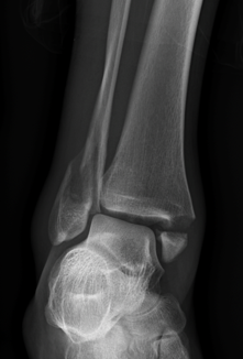
Figure 3. Mortise view x-ray of the ankle demonstrating a fracture of the distal fibula (lateral malleolus) and medial malleolus. The mortise view is taken with the foot rotated slightly inward and allows the joint space around the talus to be assessed on all sides.
How are ankle fractures treated?
Treatment options are always individually tailored. Considerations include your age, activity level, type of activity, how recently the injury occurred, which bones and ligaments are involved, how displaced the fracture is, whether the ankle mortise is congruent, whether the skin is intact, and your overall medical health, including whether you have diabetes, use nicotine, or have circulation problems.1,2
The single most important question is whether the ankle is stable. A stable ankle fracture is one in which the talus remains properly centered in the mortise and stays there under load. These fractures can generally be treated without surgery. An unstable fracture is one in which the talus can shift, and these generally do better with surgery to restore and hold the anatomy.1,2
Non-operative treatment
Non-operative treatment means allowing the fracture to heal in a cast, walking boot, or removable brace, without an operation. For the right fracture, the results are excellent, and avoiding surgery also avoids surgical risk.
The strongest evidence for this comes from the SUPER-FIN randomized trial. Investigators took 126 patients with isolated Weber B fibula fractures that appeared congruent on x-ray but were shown to be unstable on stress testing, and randomly assigned them to a cast or to surgery. At two years, the average ankle function score was 89 in the cast group and 87 in the surgery group, a difference that was neither statistically nor clinically meaningful. One patient in each group had a radiographic non-union. In the surgery group, one patient had a superficial wound infection, one had delayed wound healing, and nine required a second operation to remove hardware, two of whom developed infections afterward. Cast treatment produced fewer treatment-related harms overall.14
A meta-analysis of isolated Weber B fractures reached a similar conclusion. Surgery and non-operative care produced no significant difference in most functional scores or in return to work. Surgery produced a slightly lower AOFAS score and a slightly higher pain score in this analysis, and importantly, the total complication rate was 32.6% in the surgical group compared with 10.0% in the non-surgical group.15 A large Swedish hospital that introduced a structured treatment algorithm to reduce unnecessary surgery for stable lateral malleolar fractures found a significant reduction in reoperations and no increase in late surgeries for non-union.16
Weber A fractures, which sit below the syndesmosis, are usually stable and are almost always treated without surgery. Outcomes are generally excellent, with an average outcome score of 83 out of 100. Patients in one series reported returning to desk-based work at an average of 3.1 weeks and to labor-intensive work at 5.7 weeks, returning to physical activity at 10.8 weeks, and becoming symptom-free at 14.8 weeks. Notably, 21% of patients were still reporting some symptoms at final follow-up, so these fractures should not be dismissed as trivial.17 A recent study using CT to subclassify Weber A fractures found that when the medial soft tissues were also injured, the union rate fell from 98% to 83% and pain was more common, which suggests some Weber A fractures carry hidden instability.18
The type of immobilization also matters less than many patients expect. Comparisons of cast immobilization against removable braces show broadly similar functional outcomes.20 A randomized trial in conservatively treated Weber B fractures found that allowing early weight bearing was safe,21 and in elderly patients with more complex fracture patterns, cast treatment with early weight bearing produced acceptable quality of life and complication rates.22 In general, non-operative treatment involves 6 weeks in a cast or boot, with the bone typically healed by around 6 weeks, though full return to normal activity takes considerably longer.23
Surgical treatment
Surgery is recommended when the ankle is unstable, when the fracture is displaced and cannot be held in a good position, when the mortise is not congruent, when the ankle has dislocated, or when the fracture is open.1,2 The goal of surgery is to restore the normal anatomy of the mortise and hold it securely enough that the ankle can begin moving early.
The surgery is performed under a general anesthetic, typically with the addition of a regional nerve block.24 Nerve blocks meaningfully reduce pain medication requirements.25 Most ankle fracture surgery can be performed safely as an outpatient procedure in an ambulatory surgical center.27
For the lateral malleolus, the most common technique is a plate applied to the fibula with screws. An alternative is an intramedullary nail placed down the inside of the fibula through a much smaller incision. A meta-analysis of seven randomized trials found the infection rate with nailing was significantly lower than with plating, with no differences in function, union, or reoperation.30 Other analyses have reported similar or better functional results with nailing.31,32 This option is particularly attractive in patients with thin skin, diabetes, or poor circulation.
For the medial malleolus, fixation is usually achieved with one or two screws, sometimes with a small plate or a tension band. A randomized trial comparing percutaneous screw placement with a traditional open approach found both effective.33 Not every medial malleolus fracture requires fixation, and the decision depends on fragment size and whether the talus is displaced medially.34
For the posterior malleolus, a systematic review found that formally reducing and fixing the fragment through an incision produced significantly better patient-rated outcomes than either fixing it indirectly with screws from the front or leaving it alone.35 The size and shape of the fragment influence the decision.36 Fixation can be achieved either with screws directed from front to back or with a small plate placed through an incision at the back of the ankle; both approaches produce comparable clinical results.37
If the syndesmosis is unstable, it must be reduced and held. This is done with a flexible suture-button device. An umbrella review of nineteen systematic reviews found that suture-button fixation often produces statistically higher functional scores than screws, but that these differences usually fall below the threshold that patients can actually perceive.38 The most important factor is not which device is used but whether the syndesmosis is accurately reduced.39
The role of repairing the deltoid ligament on the inside of the ankle remains debated. A meta-analysis of Weber B and C fractures found no significant overall improvement in AOFAS score with deltoid repair, though a benefit emerged when one outlying study was excluded.40 A separate meta-analysis reported comparable findings.41 Repair is therefore usually reserved for cases where the medial side cannot otherwise be reduced. Similarly, arthroscopy can be added at the time of fixation to inspect and treat cartilage injury, though it is not required in every case.
Timing matters. Surgery is ideally performed either within the first day or two before significant swelling develops, or after the swelling has subsided, which usually takes about one to two weeks. If the ankle is dislocated it is reduced and splinted immediately regardless of when definitive surgery is planned.2
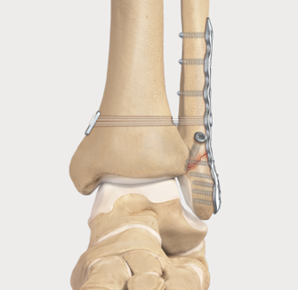
Figure 4. Fixation of a lateral malleolus (fibula) fracture with a plate and screws. In this illustration a flexible suture-button device has also been passed across the syndesmosis to hold the tibia and fibula in their normal relationship while the ligaments heal. (credit: Arthrex.com)
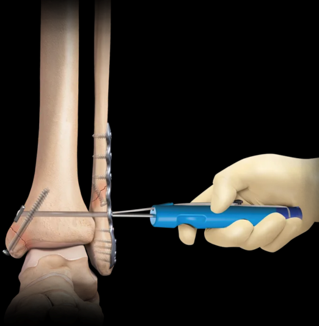
Figure 5. Fixation of a bimalleolar ankle fracture. The fibula has been stabilized with a plate and screws on the outside of the ankle, and a screw is being placed across the medial malleolus on the inside. (credit: Arthrex.com)
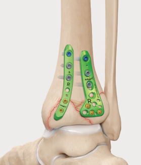
Figure 6. Fixation of a posterior malleolus fracture. Small plates placed at the back of the tibia are used to reduce and buttress the posterior fragment, an approach that has been associated with better patient-rated outcomes than leaving the fragment untreated. (credit: Arthrex.com)
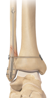
Figure 7. Intramedullary nail fixation of a distal fibula fracture. The nail is inserted down the center of the fibula through a small incision at the tip of the bone and locked in place with screws. This technique requires less soft tissue dissection than a plate and has been associated with a lower infection rate. (credit: Arthrex.com)
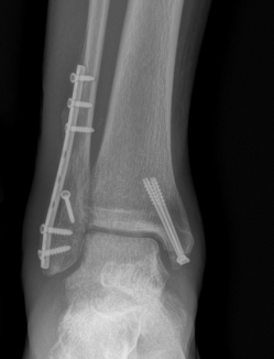
Figure 8. X-ray of an ankle after surgical fixation. A plate and screws hold the fibula on the outside of the ankle, and a screw holds the medial malleolus on the inside. The talus is centered under the tibia, indicating that the mortise has been restored.
What are the outcomes of ankle fracture surgery?
Overall, outcomes after ankle fracture surgery are good. Most patients regain a functional, largely pain-free ankle, and function improves with time, with the greatest improvement occurring during the first year.2 In one study using standard outcome instruments, nearly 90% of patients with malleolar fractures reported no pain or only mild pain, with either no limitations or limitations only in recreational activity.2
It is equally important to be realistic. Recovery from an ankle fracture is measured in months, not weeks, and a meaningful minority of patients have lasting symptoms. In another series of Weber B fractures, only 36% reported no problems at two years, and the effects of the injury extended beyond physical function into emotional wellbeing, energy, and mental health.2 Formal gait analysis confirms this: even after rehabilitation, patients who have had ankle fracture surgery walk more slowly and generate lower ankle strength than uninjured individuals, and often do not return fully to pre-injury levels.45
Several factors influence how well you do. A systematic review of 51 studies found low to moderate quality evidence that age, smoking status, and overall medical health were associated with quality of life after ankle fracture, and that age, education level, and pre-injury mobility were associated with function.46 Body weight is one of the clearest modifiable factors. In a study with up to 16 years of follow-up, obese patients had substantially worse long-term pain and function scores than non-obese patients, and pain and function declined steadily as body mass index increased. Interestingly, body mass index at the time of surgery correlated more strongly with long-term pain than body mass index at follow-up.47
Long-term results even after complex fractures can be reassuring. At an average of 11 years after a trimalleolar fracture involving the posterior malleolus, patients had mental health scores above the general population norm, and physical function scores close to normal, whether or not the posterior fragment had been fixed.48
The table below summarizes outcomes from a meta-analysis directly comparing surgical and non-surgical treatment of isolated Weber B ankle fractures. It is a useful illustration of two points: functional results are broadly similar when a fracture can reasonably be treated either way, and surgery carries additional risk that must be weighed against its benefits.
| Outcome measure | Surgical |
|---|---|
| AOFAS function score (0-100) | 91.6 |
| Olerud-Molander Ankle Score (0-100) | 83.5 |
| FAOQ score | 45.9 |
| SF-36 Physical Component Summary | 45.9 |
| SF-36 Mental Component Summary | 52.7 |
| Pain (VAS, 0-10) | 1.5 |
| Return to work at 3 months | 85.9% |
| Return to work at 6 months | 97.4% |
| Return to work at 12 months | 96.1% |
| Total complication rate | 32.6% |
| Nerve injury | 8.0% |
Table 1. Outcomes of operative versus non-operative treatment of isolated Weber B ankle fractures. Data from Tian et al, J Orthop Surg Res. 2024;19(1):346.15 Higher AOFAS and OMAS scores indicate better function; higher VAS indicates more pain. AOFAS = American Orthopaedic Foot and Ankle Society score; OMAS = Olerud-Molander Ankle Score; FAOQ = Foot and Ankle Outcome Questionnaire; PCS = Physical Component Summary; MCS = Mental Component Summary; VAS = Visual Analog Scale for pain.
When can I return to work after ankle fracture surgery?
Return to work depends far more on what your job requires than on the fracture itself, so this is a discussion we will have based on your specific occupation.
Sedentary work is usually possible relatively early, particularly if you can keep the leg elevated and have transportation. In the meta-analysis of Weber B fractures, 86% of surgically treated patients and 91% of non-surgically treated patients had returned to work by 3 months, 97% of both groups had returned by 6 months, and 96% and 99% respectively had returned by 12 months.15
How quickly you are allowed to bear weight has a direct effect on this timeline. In the INWN randomized trial, patients allowed immediate weight bearing in a walking boot after surgery returned to work significantly earlier than those kept non-weight-bearing in a cast for 6 weeks, with no increase in complications.49 A narrative review of rehabilitation trials similarly concluded that both early ankle motion and early weight bearing lead to improved function and a faster return to work.50
For work that involves prolonged standing, walking on uneven ground, climbing ladders, or heavy lifting, the timeline is longer and is usually driven by fracture healing and swelling control rather than pain. For non-operatively treated Weber A fractures, which represent the mildest end of the spectrum, patients reported returning to desk-based work at an average of 3.1 weeks and to labor-intensive work at 5.7 weeks.17 More severe fracture patterns will take substantially longer. If light duty is available, it is usually the fastest route back.
When can I return to sports after ankle fracture surgery?
Most athletes do return to sport after an ankle fracture, but the timeline varies considerably with the fracture pattern and the sport, and returning to the same level as before the injury is less certain than simply returning to activity.
A systematic review of 13 studies found return to sport rates ranging from 64% to 100%, with reported times to return ranging from about 5 weeks for an isolated lateral malleolus fracture in a professional athlete to more than 40 weeks when a syndesmotic injury was present.51 The level of sport matters a great deal. In one included study of 243 athletes, the overall return-to-sport rate was only 25%, but 88% of recreational athletes returned compared with just 12% of competitive athletes.51
A more recent study of 93 athletes treated surgically found that 83% resumed sport, with 27% returning within 3 months and 75% by one year. At one year, 41% had regained their pre-injury activity level. Fracture pattern was the strongest predictor: 100% of patients with unimalleolar fractures returned to sport at a median of 4 months, compared with 81% of those with bimalleolar fractures at 6 months and 65% of those with trimalleolar fractures at 8 months.52
There is no single test that determines readiness. In general, you should have a healed fracture on x-ray, a pain-free range of motion, and restored strength and balance before returning to cutting, pivoting, or contact sport.
| Study | Fractures (n) | Treatment | Return to sport | Time to return to sport |
|---|---|---|---|---|
| Werner et al | 237 | ORIF 128; conservative 109 | 90% | ORIF 117-124 days; conservative 72-75 days |
| Brent Lievers et al | 49 | ORIF 20; conservative 28 | 100% | Lateral malleolus 65 days; medial malleolus 38 days |
| Chiet Hong et al | 31 | ORIF 31 | 75% | NR |
| Orr et al | 72 | ORIF 72 | 64% | NR |
| Robertson et al | 96 | Conservative 52; operative 44 | 94% overall (100% conservative, 87% operative) | 26 weeks overall; 20 weeks conservative, 35 weeks operative; 43 weeks with syndesmotic injury |
| Chiet Hong et al | 47 | ORIF 47 | 81.8% (27.3% to pre-injury level) | NR |
| Colvin et al | 243 | ORIF 243 | 25% overall; 88% recreational, 12% competitive athletes | NR |
| Porter et al | 27 | ORIF 27 | 96% | Lateral malleolus 6.8 weeks; medial malleolus 17.0 weeks |
| Mai et al | 42 | ORIF 42 | 78.6% | NR |
| Walsh et al | 4 | Mixed | 100% | NR |
| Donley et al | 3 | ORIF 3 | 100% | NR |
| Navarro Garcia et al | 60 | ORIF 60 | 100% | NR |
| Pina et al | 92 | ORIF 92 | 91% | NR |
Table 2. Return to sport after ankle fracture, by study. Data from Sinha et al, Br Med Bull. 2022;143(1):35-45.51 ORIF = open reduction and internal fixation; NR = not reported.
What are the potential complications following ankle fracture surgery?
The best population-level data come from a Finnish national register of 83,666 surgically treated ankle fractures followed for up to 23 years. The overall rate of short-term complications requiring hospital readmission within 4 months was 7.2%.7 The most common long-term reoperation was removal of hardware, with a cumulative incidence of 11% at 1 year and 17% at 3 years.7 Separately, 11.5% of patients presented to an emergency department within 90 days of surgery, and 4.6% were readmitted, with most early visits related to pain or the surgical site.54
Infection
Infection is the most common complication. In the Finnish national data, infection occurred in 4.4% of cases.7 A study focused specifically on bimalleolar fractures reported comparable rates,55 and a study using a stricter definition of fracture-related infection found a prevalence of 9%.56 Risk factors include older age, diabetes, chronic kidney disease, congestive heart failure, and peripheral vascular disease.7,55,56 Prevention at the time of surgery includes sterilization of the skin, antibiotics given before incision, and careful timing of surgery relative to swelling.43 Signs of infection include fever, chills, increasing pain and swelling at the surgical site, redness, warmth, and drainage. Management may include antibiotics, surgical washout, and in some cases removal of hardware.
Blood clots
Thromboembolic complications, meaning deep vein thrombosis or pulmonary embolism, occurred in 1.6% of patients in the Finnish national data.7 A study comparing patients who did and did not receive blood thinners after ankle fracture surgery found rates of 4.1% and 3.5% respectively, with no significant difference, and no difference in wound complications or infection.57 Warning signs include new calf pain or swelling, and shortness of breath or chest pain, which should prompt immediate evaluation.
Nerve injury
Injury to the small sensory nerves around the ankle is the most common nerve-related problem and typically produces a numb patch rather than weakness. The superficial peroneal nerve, which supplies sensation to the top of the foot, can be injured in up to 21% of cases in some series.56 The sural nerve is at risk when an incision is made at the back of the ankle for a posterior malleolus fracture, and cadaveric mapping has been performed specifically to reduce this risk.60 In the Weber B meta-analysis, nerve injury occurred in 8.0% of surgically treated patients compared with 2.3% of non-surgically treated patients.15 Most of these deficits are areas of decreased sensation that improve with time.
Hardware irritation and hardware removal
Because the fibula sits just beneath thin skin, plates and screws can become irritating. This is the single most common reason for a second operation after ankle fracture surgery, with a cumulative incidence of 17% within 3 years in the Finnish national data. The risk was higher in patients under 50 and in those with bimalleolar or trimalleolar fractures.7 In a separate cohort, the most common reason cited for removal was pain around the implant, accounting for 40% of removals.61 Hardware removal is elective and is generally only considered after the fracture has fully healed.
Malunion and non-union
Malunion means the bone heals in an incorrect position; non-union means it does not heal. Both are uncommon after fixation of an ankle fracture, with mechanical complications recorded in 0.4% of cases in the Finnish register.7 When they do occur, the consequences matter, because even a small residual shift of the fibula or the talus changes how load is distributed across the joint.62 Treatment of a symptomatic malunion or non-union may involve addressing underlying causes such as nicotine use or vitamin D deficiency, use of a bone stimulator, or revision surgery to correct alignment.62
Post-traumatic arthritis
This is the most important long-term consideration. Between 75% and 80% of all ankle arthritis is post-traumatic, meaning it follows an injury.63 In a study of 477 patients followed for a minimum of 3 years after fixation, 34% had radiographic evidence of arthritis, though most were mild. Independent risk factors were age of 60 or older, heavy physical labor, a body mass index of 28 or greater, involvement of the posterior malleolus and joint surface, and residual incongruity of the joint after surgery.64 In patients under 50, the overall rate of post-traumatic arthritis was 28%, with obesity, fracture-dislocation, a large posterior malleolus fragment, and post-operative joint incongruence identified as risk factors.65 Importantly, radiographic arthritis and symptomatic arthritis are not the same thing, and many patients with x-ray changes function well.
Complications related to diabetes and nicotine
In a meta-analysis, the overall complication risk after an ankle fracture was roughly twice as high in patients with diabetes, and this risk was substantially higher with surgical than non-surgical treatment. The risk of infection was more than three times higher, and in patients with advanced diabetes the complication rate was more than eight times higher.66,67 In patients with diabetic neuropathy, the risk is higher still and treatment strategies are modified accordingly.68 Below-the-knee amputation after ankle fracture surgery is rare, with a cumulative incidence of 0.3% at maximum follow-up in the Finnish register,7 but the risk is concentrated in patients with peripheral vascular disease and chronic diabetes complications.68
Nicotine in any form is harmful to healing. Patients using non-tobacco nicotine products such as vapes had a 2.4-fold increased risk of infection and a 2.6-fold increased risk of wound problems at 30 days, and their risk of non-union remained nearly twice as high at both 1 and 2 years after surgery.69 Tobacco use carries similar risks.70
Other reported complications
Other complications include ankle stiffness, persistent swelling, wound healing problems, loss of fixation, complex regional pain syndrome, and ongoing pain. Overall mortality within 4 months of ankle fracture surgery was 0.6% in the Finnish national data, concentrated in older patients with significant medical comorbidities.7 This is not an exhaustive list. Other complications, although uncommon, can still occur.
| Complication or reoperation | Rate |
|---|---|
| Any short-term complication requiring readmission (within 4 months) | 7.2% |
| Infection | 4.4% |
| Thromboembolic complication (DVT or pulmonary embolism) | 1.6% |
| Mechanical complication | 0.4% |
| Other complications | 0.9% |
| Mortality within 4 months | 0.6% |
| Implant (hardware) removal at 1 year | 11% |
| Implant (hardware) removal at 3 years | 17% |
| Repeat fixation (osteosynthesis) at 3 years | 0.9% |
| Ankle arthroscopy at 3 years | 0.3% |
| Ankle arthrodesis (fusion) at 3 years | 0.5% |
| Below-the-knee amputation at 3 years | 0.08% |
| Ankle joint replacement at 3 years | 0.05% |
Table 3. Complications and reoperations after ankle fracture surgery in a Finnish nationwide cohort of 83,666 operatively treated ankle fractures, 1998-2020. Data from Happonen et al, J Bone Joint Surg Am. 2024;106(13):1212-1219.7
When can I drive after surgery?
There is no definitive test we can perform to determine when a patient is safe to return to driving, and this remains an area where recommendations vary widely. A systematic review of the literature on return to driving after lower-extremity surgery concluded that recommendations after a right ankle fracture range from at least 6 to 12 weeks.71
More recent work suggests that many patients are ready earlier than that. In a study that put patients through both a driving simulator and a supervised on-road driving test, 91% of patients with an isolated, surgically treated right ankle fracture passed a standard on-road driving test at 6 weeks after surgery, before their fractures had healed and before weight bearing had even been started.72 A separate study measuring braking reaction time across a range of lower-extremity injuries found that ankle and foot injuries produced the longest delays in braking, which underscores why this should not be rushed.73
Practically, several conditions should be met before you drive. You should not be taking narcotic or other sedating medications. You should be out of a cast or boot on the driving foot. You should have enough strength, motion, and reaction speed to perform an emergency stop, not just normal driving. If your left ankle was injured and you drive an automatic transmission, you may be able to return considerably earlier.
What does rehab look like after surgery?
Rehabilitation after ankle fracture surgery has changed substantially over the past decade. The traditional approach of 6 weeks of non-weight-bearing in a cast has largely given way to earlier motion and earlier weight bearing, provided the fixation is secure.
A Cochrane review of rehabilitation after ankle fracture concluded that starting weight bearing within three weeks of surgery may improve outcomes in the first six months, and that a removable ankle support during the first six weeks may also improve recovery, although in both cases the size of the benefit may be modest. Neither approach appears to increase the risk of reoperation.23 The INWN randomized trial found that patients allowed immediate weight bearing in a walking boot had significantly better ankle function at 6 weeks than those kept non-weight-bearing in a cast, along with earlier return to work, greater cost savings, and similar complication rates.49 Several meta-analyses have reached broadly consistent conclusions.74-76
Early weight bearing is not risk-free in every situation. One trial of immediate unprotected weight bearing, without a boot, found more superficial wound complications in the immediate weight-bearing group and no functional advantage at 12 weeks, which suggests protection in a boot matters.77 Laboratory work has also defined safe ranges of early passive motion so that rehabilitation does not stress the fixation.78 For this reason, your specific protocol will depend on your fracture pattern, the quality of your bone, the security of your fixation, and your medical history. Older patients, patients with diabetes or neuropathy, and patients with trimalleolar fractures are typically advanced more slowly, with recommendations extending to around 8 weeks of protected weight bearing.50
The timeline below reflects a typical protocol for a stable surgical fixation. It is a guide, not a prescription; your protocol will be individualized and may be slower or faster.
| Time after surgery | Immobilization and weight bearing | Motion and therapy | Milestones |
|---|---|---|---|
| Week 0 to 2 | Splint or walking boot. Weight bearing as directed; many stable fixations begin protected weight bearing in a boot immediately. | Elevation, toe and knee motion. No ankle motion until wound is checked. | Wound check and suture or dressing removal at first visit. Off narcotics for most patients. |
| Week 2 to 6 | Transition to removable walking boot. Progressive weight bearing as directed. | Begin active and gentle passive ankle range of motion out of the boot several times daily. Start formal physical therapy. | Improving swelling control. Most patients with sedentary jobs have returned to work. |
| Week 6 to 8 | Wean out of boot into a supportive shoe or brace once x-rays show healing. | Progressive strengthening, balance and proprioception training, calf stretching. | Radiographic union typically achieved. Driving is often possible around this point for an isolated right ankle fracture. |
| Week 8 to 12 | Full weight bearing without immobilization. | Advance strengthening, begin low-impact conditioning such as stationary cycling, swimming, and elliptical. | Return to labor-intensive work often becomes possible. Gait normalizing. |
| Month 3 to 6 | No immobilization. | Sport-specific training, agility, and impact loading as tolerated. | Return to sport for most unimalleolar fractures around 4 months; bimalleolar around 6 months. |
| Month 6 to 12 | No immobilization. | Continued strengthening and conditioning as needed. | Return to sport for trimalleolar and syndesmotic injuries. The majority of functional improvement occurs within the first year, with further gains possible beyond. |
Table 4. Representative rehabilitation timeline after surgical fixation of an ankle fracture, derived from randomized trials and systematic reviews of post-operative weight bearing and mobilization.23,49,50,52,72,74,77,79 Your individual protocol may differ.
Pain management after surgery
Narcotic pain medications and anti-inflammatories may be prescribed after surgery to help reduce pain. Nerve blocks placed at the time of surgery have been shown to significantly reduce the amount of opioid medication required, both in the recovery room and over the first 24 hours.25 Elevation of the leg above the level of the heart is one of the most effective things you can do for pain in the first two weeks, because most early pain after ankle surgery is driven by swelling. Ice, applied over the dressing rather than directly on skin, is also helpful.
Post-operative instructions
Following your stay in the recovery room and when your vital signs are stable you will be discharged to your escort.
Remember, it is normal to feel a little dizzy or drowsy for several hours after surgery. This is due to the action of the medicine used during surgery.
If you do not have a post-operative appointment scheduled, please call the office as soon as possible to schedule this appointment.
Take your pain medicine as directed. Begin the pain medicine before you start getting uncomfortable, as the nerve block will wear off. If you wait to take your pain medication until the pain is severe, you will have more difficulty in controlling the pain.
If you are taking narcotic pain medication you may need a stool softener to prevent constipation. Over-the-counter medication such as Docusate or Milk of Magnesia is recommended.
Keep the operative leg elevated above the level of your heart as much as possible for the first two weeks. This is the single most important thing you can do to reduce swelling and protect the incisions.
Notify the office of any fever, chills, or temperature greater than 100.5.
Notify the office of any wound drainage.
Notify the office immediately of new calf pain or swelling, or of shortness of breath or chest pain, as these can be signs of a blood clot.
Post-surgery diet
Resume your diet as tolerated and include vegetables, fruits, and proteins (such as meats, fish, chicken, nuts, and eggs) to promote healing. Also, remember to have adequate fluid intake. It is common after surgery to lack an appetite. This may be the result of anesthesia and the medications. Proper nutrition is needed for healing. During the healing process, the body needs increased amounts of calories and protein. Eat a variety of foods to get all the calories, proteins, vitamins, and minerals you need. If you have been told to follow a specific diet, please follow it.
How should I manage my surgical site and bandages?
You will typically be in a splint or a boot after surgery. Keep the dressing clean and dry until your first follow-up appointment. Cast covers can be purchased online to aid in keeping the splint dry during showers. Do not remove the dressing or get the incisions wet before you are cleared to do so, as wound problems are the most common early complication after this surgery.
What is my activity level after surgery?
Your weight-bearing status will be specified for you at the time of surgery and depends on your fracture pattern and fixation. Many patients with a stable fixation are allowed to bear weight in a walking boot early, which has been shown to improve early function and speed return to work without increasing complications.49 Others, particularly those with trimalleolar fractures, syndesmotic injuries, diabetes, or osteoporotic bone, will be protected longer.50
Regardless of your weight-bearing status, keep the leg elevated as much as possible early on, and move your toes and knee freely. Once you are cleared, begin the range of motion program you are given. Early ankle motion has been associated with better function and a faster return to work compared with prolonged immobilization.23,50 You should move (straighten and bend) your toes at least 10 times per day within your comfort to decrease swelling and prevent stiffness.
Can I smoke following surgery?
You should not smoke, vape, or use any nicotine product after surgery, as nicotine interferes with bone and wound healing. Patients using non-tobacco nicotine products such as vapes had more than double the risk of infection and wound problems within 30 days of ankle fracture surgery, and their risk of the fracture failing to heal remained nearly twice as high at two years.69
Questions or concerns
If at any time you have questions or concerns you can either contact your surgeon's medical assistant via email or you can call the main office numbers at: (855) 624-3306. You can also use the electronic medical record's online portal to send questions.
References
- Thome AP, Backus JD, McCormick JJ. Ankle fractures and dislocations. In: Coughlin MJ, Saltzman CL, Anderson RB, eds. Coughlin and Mann's Surgery of the Foot and Ankle. 10th ed. Philadelphia, PA: Elsevier; 2023:1854-1892.
- Adams SB, Tainter DM, Taylor MA. Malleolar fractures and soft tissue injuries of the ankle. In: Browner BD, Jupiter JB, Krettek C, Anderson PA, eds. Skeletal Trauma: Basic Science, Management, and Reconstruction. 6th ed. Philadelphia, PA: Elsevier; 2020:2446-2496.
- Obionu K, Palm H, Astrup Y, Lange NB, Krogsgaard MR. A clinical guide to the medial ligaments of the ankle: anatomy, function, diagnosis of injuries and treatment of instability—a narrative review. Foot Ankle Surg. 2024;30(2):85-91. doi:10.1016/j.fas.2023.10.010
- Möller Rydberg E, Wennergren D, Stigevall C, Ekelund J, Möller M. Epidemiology of more than 50,000 ankle fractures in the Swedish Fracture Register during a period of 10 years. J Orthop Surg Res. 2023;18(1):79. doi:10.1186/s13018-023-03558-2
- Kim TG, Choi JG, Heo YM, et al. Epidemiology and characteristics of ankle and foot injuries in electric scooter-related accidents. Clin Orthop Surg. 2024;16(2):326-334. doi:10.4055/cios23312
- Vanderkarr MF, Ruppenkamp JW, Vanderkarr M, Parikh A, Holy CE, Putnam M. Incidence, costs and post-operative complications following ankle fracture—a US claims database analysis. BMC Musculoskelet Disord. 2022;23(1):1129. doi:10.1186/s12891-022-06095-x
- Happonen V, Kröger H, Sund R. Complications after ankle fracture surgery in Finland between 1998 and 2020. J Bone Joint Surg Am. 2024;106(13):1212-1219. doi:10.2106/JBJS.23.00745
- Lau BC, Allahabadi S, Palanca A, Oji DE. Understanding radiographic measurements used in foot and ankle surgery. J Am Acad Orthop Surg. 2022;30(2):e139-e154. doi:10.5435/JAAOS-D-20-00189
- Gitajn IL, Natoli RM, Spitler CA, et al. Radiographic assessment of ankle fracture displacement: a validation study. Foot Ankle Int. 2022;43(10):1269-1276. doi:10.1177/10711007221106471
- Ali RA, Mitchell W, Bora VG, Dawson JR, Atassi OH, Perkins CH. Effectiveness of gravity and manual stress radiographs and the use of lateral talar displacement in determining ankle stability of supination-external rotation type ankle fractures. J Orthop Trauma. 2021;35(12):e475-e480.
- Seo M, Kwon M, Sung KS. Weight-bearing test: a straightforward and reliable indicator of stability in supination-external rotation type, Weber B ankle fractures. Clin Orthop Surg. 2026;18(4):543-547. doi:10.4055/cios25349
- Kerut CK, Mudiganty S, Williams R, Valencia M, Kerut DG, Gonzales J. Probability of occult ankle fracture based on radiograph-measured swelling. J Am Acad Orthop Surg Glob Res Rev. 2024;8(5):e23.00271. doi:10.5435/JAAOSGlobal-D-23-00271
- Williamson ERC, Shimozono Y, Toale J, et al. Incidence of chondral and osteochondral lesions in ankle fracture patients identified with ankle arthroscopy following rotational ankle fracture: a systematic review. J Foot Ankle Surg. 2022;61(3):668-673. doi:10.1053/j.jfas.2021.12.013
- Kortekangas T, Lehtola R, Leskelä HV, et al. Cast immobilisation versus surgery for unstable lateral malleolus fractures (SUPER-FIN): randomised non-inferiority clinical trial. BMJ. 2025;390:e085295. doi:10.1136/bmj-2025-085295
- Tian J, Miao J, Jiang Z, Li Z. Comparison of operatively and nonoperatively treated isolated Weber B ankle fractures: a systematic review and meta-analysis. J Orthop Surg Res. 2024;19(1):346. doi:10.1186/s13018-024-04835-4
- Börjesson E, Johannesson K, Ekelund J, Möller Rydberg E. Non-surgical treatment of lateral malleolar fractures is safe: long-term follow-up of a comprehensive treatment algorithm. BMC Musculoskelet Disord. 2024;25(1):818. doi:10.1186/s12891-024-07924-x
- Chan O, Lee C, Titheradge R, Auld F, Iliopoulos E. Patient reported outcomes following non-operatively managed Weber Type A distal fibula fractures. Foot Ankle Surg. 2022;28(1):62-65. doi:10.1016/j.fas.2021.01.013
- Wu K, Ji Y, Yuan T, et al. Determinants of surgical necessity in Danis-Weber A lateral malleolus fractures: prognostic insights from a nonoperative cohort. Injury. 2026;57(1):113227. doi:10.1016/j.injury.2026.113227
- Carter TH, Oliver WM, Bell KR, Graham C, Duckworth AD, White TO. Operative vs nonoperative management of unstable medial malleolus fractures: a randomized clinical trial. JAMA Netw Open. 2024;7(1):e2351308. doi:10.1001/jamanetworkopen.2023.51308
- Zhou S, Zheng H, Cao M, et al. Comparative efficacy of cast immobilization versus removable braces in patients with ankle fractures: a systematic review and meta-analysis. BMC Musculoskelet Disord. 2025;26(1):243. doi:10.1186/s12891-025-08451-z
- Stassen RC, Franssen S, Meesters B, Boonen B, de Loos ER, van Vugt R. Prospective randomized controlled trial: early weight bearing after conservative treatment of Weber B ankle fractures (pancake trial). Eur J Orthop Surg Traumatol. 2024;34(1):591-598. doi:10.1007/s00590-023-03651-6
- Lorente A, Gandia A, Mariscal G, Palacios P, Lorente R. Quality of life and complications in elderly patients after pronation rotation type III ankle fractures treated with a cast and early weight-bearing. BMC Musculoskelet Disord. 2021;22(1):878. doi:10.1186/s12891-021-04745-0
- Lewis SR, Pritchard MW, Parker R, et al. Rehabilitation for ankle fractures in adults. Cochrane Database Syst Rev. 2024;9(9):CD005595. doi:10.1002/14651858.CD005595.pub4
- Sharrow CM, Elmore B. Anesthesia for the patient undergoing foot and ankle surgery. Anesthesiol Clin. 2024;42(2):263-280. doi:10.1016/j.anclin.2023.11.008
- Entezari B, Gleicher Y, Matelski J, Wolfstadt JI. The impact of peripheral nerve block on the quality of care after ankle fracture surgery: a quality improvement study. J Orthop Trauma. 2023;37(3):e111-e117.
- Quan T, Malyavko A, Ifarraguerri AM, et al. Evaluating the association of anesthesia type with postoperative complications in patients undergoing ankle fracture repair. J Foot Ankle Surg. 2022;61(5):1086-1090. doi:10.1053/j.jfas.2022.01.025
- Hollawell SM, Yancovitz S, Casciato DJ, Coleman MR. Safety and outcome measures of ankle open reduction and internal fixation in an ambulatory surgical center. J Foot Ankle Surg. 2024;63(3):376-379. doi:10.1053/j.jfas.2024.01.009
- Dhillon MS, Rajnish RK, Kumar P, Sharma S, Singh GP, Srivastava A. A comparison of outcomes of locking versus non-locking plate fixation for the distal fibula fractures: a systematic review and meta-analysis. Eur J Orthop Surg Traumatol. 2024;34(1):75-89. doi:10.1007/s00590-023-03694-9
- Hasami NA, Smeeing DPJ, Pull ter Gunne AF, Edwards MJR, Nelen SD. Operative fixation of lateral malleolus fractures with locking plates vs nonlocking plates: a systematic review and meta-analysis. Foot Ankle Int. 2022;43(2):280-290. doi:10.1177/10711007211040508
- Wang J, Jia HB, Li HM, Jiang HQ, Zhao JG. Intramedullary nailing versus open reduction and plate fixation for lateral malleolar fractures: a meta-analysis. J Foot Ankle Surg. 2024;63(2):275-280. doi:10.1053/j.jfas.2023.11.016
- Attia AK, Fayed A, Mahmoud K, Labib SA, Aydogan U, Juliano P. Locked intramedullary nailing provides superior functional outcomes and lower complication rates than plate fixation of distal fibula fractures: a systematic review and meta-analysis of comparative studies. Foot Ankle Surg. 2022;28(7):986-994. doi:10.1016/j.fas.2022.02.005
- Rushing CJ, Spinner SM, Armstrong AV, Hardigan PC. Intramedullary fixation of distal fibula fractures. Foot Ankle Spec. 2021;14(2):104-112. doi:10.1177/1938640021991735
- Ahmed KFE. Percutaneous versus open cannulated screws fixation for displaced isolated medial malleolar fractures in adults: a randomized controlled clinical trial. Arch Orthop Trauma Surg. 2025;145(1):385. doi:10.1007/s00402-025-06000-w
- Pinski JM, Ryan SP, Pittman JL, Tornetta P. Is fixation of the medial malleolus necessary in unstable ankle fractures? Arch Orthop Trauma Surg. 2023;143(6):2999-3005. doi:10.1007/s00402-022-04528-9
- Miksch RC, Herterich V, Barg A, Böcker W, Polzer H, Baumbach SF. Open reduction and internal fixation of the posterior malleolus fragment in ankle fractures improves the patient-rated outcome: a systematic review. Foot Ankle Int. 2023;44(8):727-737. doi:10.1177/10711007231165771
- Patel S, Baburaj V, Sharma S, Mason LW, Dhillon MS. Influence of posterior malleolar fragment morphology on the outcomes of trimalleolar fractures: a systematic review and meta-analysis. Foot Ankle Surg. 2022;28(8):1337-1344. doi:10.1016/j.fas.2022.06.012
- Gao Y, Liu Y, Zhao Y, et al. Comparison between anterior-to-posterior screw fixation versus posterolateral approach plate fixation for posterior malleolus fracture: a systematic review and meta-analysis. Foot Ankle Surg. 2024;30(7):594-602. doi:10.1016/j.fas.2024.05.004
- Nieuwenkamp CL, Tausendfreund J, Schepers T. Suture button versus syndesmotic screw fixation in acute ankle fractures with syndesmotic injury: an umbrella review of functional outcomes and clinical relevance based on the minimal clinically important difference. Injury. 2026;57(1):113054. doi:10.1016/j.injury.2026.113054
- Kaiser PB, Bejarano-Pineda L, Kwon JY, DiGiovanni CW, Guss D. The syndesmosis, part II: surgical treatment strategies. Orthop Clin North Am. 2021;52(4):417-432. doi:10.1016/j.ocl.2021.05.011
- D'Andrea G, Colombo P, Placella G, Salini V, Alessio-Mazzola M. The role of deltoid ligament repair in Weber B and C ankle fractures: a systematic review and meta-analysis of clinical, functional and radiographic results. J Foot Ankle Surg. 2025;64(6):812-819. doi:10.1053/j.jfas.2025.06.010
- Guo W, Lin W, Chen W, Pan Y, Zhuang R. Comparison of deltoid ligament repair and non-repair in acute ankle fracture: a meta-analysis of comparative studies. PLoS One. 2021;16(11):e0258785. doi:10.1371/journal.pone.0258785
- Miksch RC, Spindler FT, Böcker W, Polzer H, Baumbach SF. Arthroscopically assisted (AORIF) ankle fracture treatment seems to lead to superior results when compared to open reduction and internal fixation (ORIF) only: results of a systematic review. Arch Orthop Trauma Surg. 2025;145(1):451. doi:10.1007/s00402-025-06030-4
- Sugino T, Takegami Y, Bando K, et al. The wait time for surgery following injury affects functional outcomes and complications after an ankle fracture: a propensity score-matched multicenter study, the TRON study. Foot Ankle Spec. 2024;17(6):592-600. doi:10.1177/19386400231164211
- Hawkins J, Andrews NA, Sankey MT, et al. The impact of surgical timing after ankle fracture on clinical and long-term patient reported outcomes. J Foot Ankle Surg. 2023;62(4):701-706. doi:10.1053/j.jfas.2023.02.011
- Long Q, Yang Z, Shao T, Liu Y. Comparison of gait parameters between post-surgery ankle fracture patients and healthy individuals: a systematic review and meta-review. Biomed Eng Online. 2025;24(1):128. doi:10.1186/s12938-025-01463-9
- Chan BHT, Snowdon DA, Williams CM. The association between person and fracture characteristics with patient reported outcome after ankle fractures in adults: a systematic review. Injury. 2022;53(7):2340-2365. doi:10.1016/j.injury.2022.02.016
- Cardoso DV, Paccaud J, Dubois-Ferrière V, et al. The effect of BMI on long-term outcomes after operatively treated ankle fractures: a study with up to 16 years of follow-up. BMC Musculoskelet Disord. 2022;23(1):317. doi:10.1186/s12891-022-05247-3
- Chidda A, Soares S, Tannast M, Schwab J, Seidel A. Long-term outcomes after a trimalleolar fracture involving the posterior malleolar fragment: an 11-year follow-up. Arch Orthop Trauma Surg. 2025;145(1):346. doi:10.1007/s00402-025-05959-w
- Khojaly R, Rowan FE, Shah V, et al. Immediate weight-bearing compared with non-weight-bearing after operative ankle fracture fixation: results of the INWN pragmatic, randomized, multicenter trial. J Bone Joint Surg Am. 2025;107(13):1423-1434. doi:10.2106/JBJS.24.00965
- Jafarian Tangrood Z, Hooper G, Mowbray J, Halai M, Powell A. Rehabilitation protocols after ankle surgeries: a narrative literature review. Trauma. 2025;27(4):265-272. doi:10.1177/14604086251352075
- Sinha A, Robertson G, Maffulli N. Doctor, I fractured my ankle. When can I return to play? An updated systematic review. Br Med Bull. 2022;143(1):35-45. doi:10.1093/bmb/ldac016
- Saliba I, Cannell S, Fontanier V, et al. Predictive factors to return to sport after surgical management of ankle fractures. J Foot Ankle Surg. 2025;64(2):197-204. doi:10.1053/j.jfas.2024.10.003
- Saliba I, Cannell S, Valentin E, et al. Validation of the Ankle Ligament Reconstruction-Return to Sports after Injury (ALR-RSI) score as a tool to assess psychological readiness to return to sport in an active population after ankle fracture surgery: a cross-sectional study. J Foot Ankle Surg. 2024;63(3):295-299. doi:10.1053/j.jfas.2023.12.005
- Kammien AJ, Ratnasamy PP, Joo PY, et al. Ninety-day emergency department visits after ankle fracture surgery. J Am Acad Orthop Surg. 2023;31(1):e51-e57. doi:10.5435/JAAOS-D-22-00484
- Mastrokostas PG, Monas A, Rodriguez AN, Lam A, et al. Patient demographics and risk factors for surgical site infections after open reduction and internal fixation for bimalleolar ankle fractures. J Am Acad Orthop Surg. 2026;34(14):e1907-e1914. doi:10.5435/JAAOS-D-24-01181
- Pilskog K, Høvding P, Fenstad AM, Inderhaug E, Fevang JM, Dale H. Risk factors for fracture-related infection after ankle fracture surgery. Injury. 2023;54(12):111011. doi:10.1016/j.injury.2023.111011
- Elliott IS, Rane AA, DeKeyser GJ, et al. Venous thromboembolism in patients with surgically treated ankle fractures. Arch Orthop Trauma Surg. 2023;143(3):1237-1242. doi:10.1007/s00402-021-04192-5
- Brennan J, Keblish D, Friedmann E, Spirt A, Holt E, Turcotte J. Postoperative venous thromboembolism risk-prediction in foot and ankle fracture surgery. Foot (Edinb). 2023;56:102017. doi:10.1016/j.foot.2023.102017
- Peng S, Zhang M, Jin J, MacCormick AD. The effectiveness of venous thromboembolism prophylaxis interventions in trauma patients: a systematic review and network meta-analysis. Injury. 2023;54(12):111078. doi:10.1016/j.injury.2023.111078
- Ghani Y, Najefi AA, Aljabi Y, Vemulapalli K. Anatomy of the sural nerve in the posterolateral approach to the ankle: a cadaveric study. J Foot Ankle Surg. 2023;62(2):286-290. doi:10.1053/j.jfas.2022.08.001
- Karagoz B, Erdem M, Bayrak HC, Bakir M, Agir I. Determinants of implant removal in isolated Weber type B and C ankle fractures: a retrospective cohort study. J Foot Ankle Surg. 2026;65(1):46.e1-46.e5. doi:10.1053/j.jfas.2025.10.004
- Ingall EM, Zhao J, Kwon JY. Revision strategies for the aseptic, malaligned, surgically treated ankle fracture. Foot Ankle Clin. 2022;27(2):355-370. doi:10.1016/j.fcl.2021.11.022
- Anastasio AT, Lau B, Adams S. Ankle osteoarthritis. J Am Acad Orthop Surg. 2024;32(16):738-746. doi:10.5435/JAAOS-D-23-00743
- Wang L, Ji C, Zhang X, et al. Post-traumatic osteoarthritis after ankle fracture fixation: a minimum three-year follow-up. Foot Ankle Surg. 2025;31(5):576-582. doi:10.1016/j.fas.2025.02.014
- Beak JS, Kim YT, Lee SH. Predisposing factors for posttraumatic osteoarthritis after malleolus fracture fixation in patients younger than 50 years. Foot Ankle Int. 2022;43(3):389-397. doi:10.1177/10711007211050039
- Lopez-Capdevila L, Rios-Ruh JM, Fortuño J, et al. Diabetic ankle fracture complications: a meta-analysis. Foot Ankle Surg. 2021;27(8):832-837. doi:10.1016/j.fas.2020.10.015
- Amini B, Kadhm S, Trompeter A. The impact of diabetes mellitus on the management and outcome of ankle fractures. Injury. 2025;56(4):112226. doi:10.1016/j.injury.2025.112226
- Kirchner GJ, Kim AH, Martinazzi BJ, Sudah SY, Lieber AM, Aynardi MC. Factors associated with amputation following ankle fracture surgery. J Foot Ankle Surg. 2023;62(5):792-796. doi:10.1053/j.jfas.2023.04.002
- Sontam TR, Toussaint L, Hlavinka WJ, Chavda H, Mbilinyi RH, Zide JR. Elevated risk of postoperative complications with non-tobacco nicotine use following open reduction and internal fixation of ankle fractures. J Am Acad Orthop Surg. 2026;34(14):e1927-e1934. doi:10.5435/JAAOS-D-25-00714
- Dhodapkar MM, Oghenesume OP, Halperin SJ, Modrak M, Yoo BJ, Grauer JN. Adverse events after ankle fracture open reduction internal fixation among patients with and without documented cannabis and tobacco use. Foot Ankle Int. 2023;44(10):941-948. doi:10.1177/10711007231189698
- Frane N, Bandovic I, Hu V, Bitterman A. Return-to-driving recommendations after lower-extremity orthopaedic procedures. JBJS Rev. 2020;8(12):e20.00066. doi:10.2106/JBJS.RVW.20.00066
- Ho SWL, Yam M, Chan ML, Kwek EBK. Return to car driving is safe 6 weeks after operative treatment of right ankle fractures. Arch Orthop Trauma Surg. 2018;138(12):1691-1697. doi:10.1007/s00402-018-3037-3
- Alejo AL, Rascoe A, Kim CY, et al. Prolonged time to brake following lower extremity injuries. J Am Acad Orthop Surg Glob Res Rev. 2023;7(4):e23.00018. doi:10.5435/JAAOSGlobal-D-23-00018
- Sharma T, Farrugia P. Early versus late weight bearing & ankle mobilization in the postoperative management of ankle fractures: a systematic review and meta-analysis of randomized controlled trials. Foot Ankle Surg. 2022;28(7):827-835. doi:10.1016/j.fas.2022.03.003
- Tong J, Ajrawat P, Chahal J, et al. Early versus delayed weight bearing and mobilization after ankle fracture fixation surgery: a systematic review and meta-analysis of randomized controlled trials. Orthopedics. 2024;47(1):e1-e9. doi:10.3928/01477447-20230804-08
- Sernandez H, Riehl J, Fogel J. Do early weight-bearing and range of motion affect outcomes in operatively treated ankle fractures: a systematic review and meta-analysis. J Orthop Trauma. 2021;35(8):408-413.
- Le V, Viskontas D, Lohre R, et al. Immediate unprotected weightbearing vs 2 weeks nonweightbearing after open reduction internal fixation of ankle fractures. Foot Ankle Int. 2024;45(2):103-114. doi:10.1177/10711007231217675
- Ni M, Sun T, Zhang T, Jin J, Song Y. Quantitative initial safety range of early passive rehabilitation after ankle fracture surgery. Injury. 2022;53(6):2281-2286. doi:10.1016/j.injury.2022.03.067
- Jarragh A, AlAwadhi K, Shammasi A, et al. Splint versus no splint after ankle fracture fixation; results from the multi-centre post-operative ankle splint trial (PAST). Injury. 2023;54(12):111084. doi:10.1016/j.injury.2023.111084
- Agarwalla A, Gowd AK, Cody EA, Tan EW, et al. Prediction of short-term postoperative complications following open reduction internal fixation of ankle fractures. J Am Acad Orthop Surg. 2026;34(14):e2000-e2011. doi:10.5435/JAAOS-D-25-00532
- Aragoneses López P, López Hernández G, Malagelada F. The role of arthroscopy in ankle fracture fixation. Foot Ankle Clin. 2025;30(4):723-736. doi:10.1016/j.fcl.2025.08.001
- Attia AK, Urdahl TH, Dock CC, et al. Outcomes of deltoid ligament repair in surgically treated Weber C ankle fractures with deltoid injury: a retrospective comparative study. Foot Ankle Int. 2025;46(10):1139-1146. doi:10.1177/10711007251361122
- Bando K, Takegami Y, Ando T, et al. Early weight bearing and mobilization decrease perioperative complications in patients after ankle fracture: the retrospective multicenter (TRON group) study. J Orthop Sci. 2023;28(3):651-655. doi:10.1016/j.jos.2022.03.002
- Bartoníček J, Rammelt S, Tuček M. Maisonneuve fractures of the ankle: a critical analysis review. JBJS Rev. 2022;10(2):e21.00160. doi:10.2106/JBJS.RVW.21.00160
- Bayrak HC, Karagoz B, Gök O. Clinical and radiological outcomes of screw fixation and suture-button technique in syndesmosis injuries with isolated lateral malleolus fractures. J Foot Ankle Surg. 2026;65(1):56.e1-56.e9. doi:10.1053/j.jfas.2025.11.007
- Bejarano-Pineda L, DiGiovanni CW, Guss D, Waryasz G. Diagnosis and treatment of syndesmotic unstable injuries: where we are now and where we are headed. J Am Acad Orthop Surg. 2021;29(23):985-997. doi:10.5435/JAAOS-D-20-01350
- Bejarano-Pineda L, Guss D, Waryasz G, DiGiovanni CW, Kwon JY. The syndesmosis, part I: anatomy, injury mechanism, classification, and diagnosis. Orthop Clin North Am. 2021;52(4):403-415. doi:10.1016/j.ocl.2021.05.010
- Bretherton CP, Kotwal T, Sandhu H, Baird J, Griffin XL. Patient experiences and desires for recovery after ankle fracture surgery. Injury. 2024;55(11):111763. doi:10.1016/j.injury.2024.111763
- Chen B, Ye Z, Wu J, Wang G, Yu T. The effect of early weight-bearing and later weight-bearing rehabilitation interventions on outcomes after ankle fracture surgery: a systematic review and meta-analysis of randomised controlled trials. J Foot Ankle Res. 2024;17(2):e12011. doi:10.1002/jfa2.12011
- Chong LSL, Khademi M, Reddy KM, Anderson GH. Ten year outcomes after non-fixation of the smaller posterior malleolar fragment: a retrospective cohort study. Foot (Edinb). 2024;59:102091. doi:10.1016/j.foot.2024.102091
- Davey MS, Davey MG, Hurley ET, Kearns SR. Tourniquet use during open reduction and internal fixation of ankle fractures—a systematic review and meta-analysis. J Foot Ankle Surg. 2022;61(5):1103-1108. doi:10.1053/j.jfas.2022.01.019
- Fischer FS, Muhammad M, Wagner RK, et al. Patient-reported and radiographic outcomes following operative fixation of bimalleolar equivalent ankle fractures with deltoid ligament repair. J Foot Ankle Surg. 2026;65(2):111.e1-111.e5. doi:10.1053/j.jfas.2026.01.015
- Fűzy EJ, Ferreira N, Brown C, Hugo D, Joubert E, Burger M. The reliability of posterior malleolar ankle fracture assessment: a unique perspective. Eur J Orthop Surg Traumatol. 2024;34(1):539-548. doi:10.1007/s00590-023-03702-y
- Gopinath R, Pigott M, Lindsey B, et al. Medial ankle instability: review of anatomy, evaluation, and treatment. Foot Ankle Spec. 2022;15(6):574-583. doi:10.1177/1938640021992915
- Guedes S, Sousa-Pinto B, Torres J. Radiological outcomes of bimalleolar fractures: are timing of surgery and type of reconstruction important? Orthop Traumatol Surg Res. 2022;108(5):103314. doi:10.1016/j.otsr.2022.103314
- Guo W, Wu F, Chen W, Tian K, Zhuang R, Pan Y. Can locked fibula nail replace plate fixation for treatment of acute ankle fracture? A systematic review and meta-analysis. J Foot Ankle Surg. 2023;62(1):178-185. doi:10.1053/j.jfas.2022.10.003
- Gwak HC, Kim JH, Park DH, et al. Patterns of anterior inferior tibiofibular ligament avulsion fracture accompanied by ankle fracture. J Foot Ankle Surg. 2024;63(1):127-131. doi:10.1053/j.jfas.2023.06.006
- Halai M, Meulenkamp B, Buckley R. Should a significantly displaced ankle fracture have an ankle arthroscopy before it is treated with ORIF? Injury. 2023;54(2):791-793. doi:10.1016/j.injury.2022.11.044
- Hegde A, Shetty C, Joseph N, Mane P. Comparison of posterior and lateral surface plating for type B Weber lateral malleoli fractures. Foot Ankle Spec. 2024;17(2):130-138. doi:10.1177/19386400221079197
- Heyes GJ, Khashkhusha TR, Mason L. Posterolateral approach to posterior malleolar fractures: a literature review. Foot Ankle Spec. 2023;16(2):176-184. doi:10.1177/19386400211009366
- Hollawell SM, Abergel P, Yancovitz S, Casciato DJ, Coleman MR. Comparison of clinical outcomes in acute ankle fracture open reduction internal fixation with and without ankle arthroscopy. J Foot Ankle Surg. 2025;64(6):767-772. doi:10.1053/j.jfas.2025.06.006
- Hunt AA, Maschhoff C, Van Rysselberghe N, Gonzalez CA, Goodnough H, Gardner M, Bishop JA. Historic indications for fixation of posterior malleolus fractures—where did they come from and where are we now? Injury. 2024;55(6):111537. doi:10.1016/j.injury.2024.111537
- James M, Dodd A. Management of deltoid ligament injuries in acute ankle fracture: a systematic review. Can J Surg. 2022;65(1):E9-E17. doi:10.1503/cjs.020320
- Jupiter DC, Hsu ES, Liu GT, Reilly JG, Shibuya N. Risk factors for short-term complication after open reduction and internal fixation of ankle fractures: analysis of a large insurance claims database. J Foot Ankle Surg. 2020;59(2):239-245. doi:10.1053/j.jfas.2019.08.003
- Karaismailoglu B, Yıldırım O, Aslan L, Yontar NS, Guven MF, Kaynak G, Ogut T. Middle-sized posterior malleolus fractures: fixed versus non-fixed—a prospective randomized study. Foot Ankle Surg. 2023;29(4):329-333. doi:10.1016/j.fas.2023.04.005
- Khojaly R, Rowan FE, Hassan M, Hanna SA, Mac Niocaill R. Weight-bearing allowed following internal fixation of ankle fractures, a systematic literature review and meta-analysis. Foot Ankle Int. 2022;43(9):1143-1156. doi:10.1177/10711007221102142
- Kim SH, Lee SH, Lee YK. The clinical outcomes of syndesmotic flexible fixation for syndesmotic injury with ankle fracture. Foot Ankle Int. 2023;44(11):1112-1119. doi:10.1177/10711007231194045
- Kohler FC, Schenk P, Koehler P, et al. The role of the posterior malleolus in the treatment of unstable upper ankle joint injuries—a biomechanical study. Foot Ankle Surg. 2022;28(7):979-985. doi:10.1016/j.fas.2022.02.006
- Kohli M, Atodaria KP, Patel R, Brahmabhatt S, Goyal AR, Raval N. Incidence of deep vein thrombosis and pulmonary embolism in closed foot and ankle fractures treated with conservative versus surgical management: a nationwide analysis. Injury. 2024;55(11):111859. doi:10.1016/j.injury.2024.111859
- Lam A, Tabbaa A, Rodriguez A, et al. Early full weight-bearing is safe following syndesmotic injury: a prospective randomized comparative study. Foot Ankle Orthop. 2025;10(1):2473011425S00222. doi:10.1177/2473011425S00222
- Lim B, Shaalan M, O'hEireamhoin S, Lyons F. Syndesmotic fixation in Weber B ankle fractures: a systematic review. PLoS One. 2024;19(6):e0304148. doi:10.1371/journal.pone.0304148
- Liu G, Sun Z, Yang M, Wu X, Giannoudis PV. An overview on the management of ankle fractures in elderly patients aged 65 and over: a scoping review. Eur J Orthop Surg Traumatol. 2025;35(1):396. doi:10.1007/s00590-025-04512-0
- Loubignac F. Treatment of bimalleolar fractures in elderly. Orthop Traumatol Surg Res. 2022;108(1):103137. doi:10.1016/j.otsr.2021.103137
- Oliver W, Ha J, Buckley R. Should all unstable ankle fractures with a small posterior malleolar fracture have lateral, syndesmotic and posterior malleolar fixation? Injury. 2025;56(5):112453. doi:10.1016/j.injury.2025.112453
- Phyo N, Wee A. Managing acute ankle and hindfoot fracture in diabetic patients. Foot Ankle Clin. 2022;27(3):639-654. doi:10.1016/j.fcl.2022.02.002
- Pilskog K, Gote TB, Odland HEJ, et al. Association of delayed surgery for ankle fractures and patient-reported outcomes. Foot Ankle Int. 2022;43(6):762-771. doi:10.1177/10711007211070540
- Pinzur MS. Unstable ankle fractures in neuropathic diabetics. J Am Acad Orthop Surg. 2022;30(23):1116-1122. doi:10.5435/JAAOS-D-22-00375
- Ramsodit KR, Gouttebarge V, et al. Most elite athletes return to preinjury competitive activity after surgical treatment for medial malleolus stress fractures. Knee Surg Sports Traumatol Arthrosc. 2024;32(9):2280-2290. doi:10.1002/ksa.12284
- Rigby RB, Fleisig GS, Waldrop NE 3rd. Primary deltoid repair for ankle fracture using all-suture anchors. J Foot Ankle Surg. 2023;62(4):723-726. doi:10.1053/j.jfas.2023.03.002
- Rushing CJ. Comparison of ankle fracture fixation using intramedullary fibular nailing versus plate fixation. J Foot Ankle Surg. 2024;63(5):546-556. doi:10.1053/j.jfas.2024.05.004
- Santolini E, Giordano V, Giannoudis PV. Effect of mechanical stability of osteosynthesis on infection rates: timing of temporary and definitive fixation. Injury. 2024;55(11):111845. doi:10.1016/j.injury.2024.111845
- Schlickewei C, Krähenbühl N, Rolvien T, et al. Surgical outcome of avulsion fractures of the distal fibula: a systematic literature review. Arch Orthop Trauma Surg. 2023;143(3):1275-1291. doi:10.1007/s00402-021-04256-6
- Seo J, Yang KH, Shim DW, Cho H, Park YC. Marginal impaction associated with posterior malleolar fracture in rotational ankle injury. Injury. 2022;53(2):756-761. doi:10.1016/j.injury.2021.12.013
- Stringfellow TD, Coffey D, Wek C, et al. Epidemiology & management of complex ankle fractures in the United Kingdom: a multicentre cohort study. Injury. 2024;55(2):111037. doi:10.1016/j.injury.2023.111037
- Timing of fracture fixation in ankle fracture-dislocations. Foot Ankle Spec. 2024;17(4):368-376. doi:10.1177/19386400241273105
- Toru HK, Khan AA, Ali N. Operative vs. nonoperative management of isolated Weber B ankle fractures. Cureus. 2025;17(1):e78028. doi:10.7759/cureus.78028
- Walsh JP, Hsiao MS, LeCavalier D, McDermott R, Gupta S, Watson TS. Clinical outcomes in the surgical management of ankle fractures: a systematic review and meta-analysis of fibular intramedullary nail fixation vs. open reduction and internal fixation in randomized controlled trials. Foot Ankle Surg. 2022;28(7):836-844. doi:10.1016/j.fas.2022.03.009
- Wang C, Li C. Early weight-bearing after ankle fracture surgery: a systematic review and meta-analysis of functional outcomes and safety. J Orthop Surg Res. 2025;20(1):785. doi:10.1186/s13018-025-06216-x
- Wang J, Jia HB, Zhao JG, Wang J, Zeng XT. Plate versus screws fixation for the posterior malleolar fragment in trimalleolar ankle fractures. Injury. 2023;54(2):761-767. doi:10.1016/j.injury.2022.10.032
- Xu Y, Kang R, Li M, et al. The clinical efficacy of suture-button fixation and trans-syndesmotic screw fixation in the treatment of ankle fracture combined with distal tibiofibular syndesmosis injury: a retrospective study. J Foot Ankle Surg. 2022;61(1):143-148. doi:10.1053/j.jfas.2021.07.009
- van Halsema MS, Boers RAR, Leferink VJM. An overview on the treatment and outcome factors of ankle fractures in elderly men and women aged 80 and over: a systematic review. Arch Orthop Trauma Surg. 2022;142(11):3311-3325. doi:10.1007/s00402-021-04161-y
If you would like to have additional information on the treatment of ankle fractures, please contact Arturo Villarreal M.D., Board Certified Orthopaedic Surgeon, serving the communities of Georgetown, Cedar Park, Round Rock, TX.


