
- Rotator Cuff Pain
- Rotator Cuff Arthropathy
- Partial Rotator Cuff Tear
- Rotator Cuff Calcification
- Rotator Cuff Bursitis
What is the rotator cuff?
The rotator cuff is a group of four muscles and their tendons that surround the ball-and-socket joint of the shoulder. The four muscles are the supraspinatus, infraspinatus, teres minor and subscapularis. They begin on the shoulder blade, and their tendons attach onto the top of the arm bone (the humerus).1
The shoulder is the most mobile joint in the body, and that mobility comes at the cost of bony stability. The socket is shallow, so the shoulder depends on soft tissue rather than bone to stay centered. This is the rotator cuff's main job. Working together, the cuff muscles pull the ball of the humerus into the socket and hold it there while the much larger deltoid muscle lifts the arm. The front and back portions of the cuff - the subscapularis in front and the infraspinatus and teres minor behind - work as a balanced pair, sometimes called a force couple.2
The cuff also rotates the arm. The subscapularis rotates the arm inward, the infraspinatus and teres minor rotate it outward, and the supraspinatus helps lift the arm away from the body.3
Two other structures deserve mention because they come up frequently in discussions of rotator cuff surgery. The first is the superior capsule, a layer of tissue on the undersurface of the cuff tendons that resists upward migration of the ball. The second is the subacromial bursa, a fluid-filled sac that sits between the rotator cuff and the bone above it. The bursa allows the tendons to glide smoothly, and when it becomes inflamed it is a significant source of shoulder pain.5
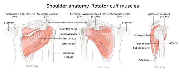
Figure 1. The muscles of the rotator cuff — the supraspinatus, infraspinatus, teres minor and subscapularis — shown from the back, front and side. (Licensed from Adobe Stock.)
How is the rotator cuff injured?
Most rotator cuff tears are degenerative. They develop gradually over years as the tendon wears out, rather than from a single injury. Laboratory studies of human cuff tendons show that the tissue becomes measurably weaker and structurally disorganized with age, and that repeated submaximal loading causes fatigue damage to accumulate within the tendon over time - much the way a paper clip bent back and forth repeatedly will eventually break.6,7
Because of this, age is by far the strongest risk factor for a rotator cuff tear. The prevalence of tears rises steadily with each decade of life, from roughly 10% of people aged 20 years or younger to more than 60% of people aged 80 years or older.8 Prevalence has been estimated to be as high as 80% after age 80.9 For many people, some degree of rotator cuff wear is simply a part of normal aging.8
Several other factors have been associated with rotator cuff disease:
- Tobacco use is associated with rotator cuff disease and, as discussed later in this guide, with poorer healing after repair.10
- Family history. Siblings of patients with rotator cuff tears have higher rates of both symptomatic and asymptomatic tears, and their tears are more likely to progress.11
- Medical conditions. Diabetes, high blood pressure and high cholesterol have each been associated with rotator cuff disease.12
- Shoulder shape. The shape and slope of the acromion - the shelf of bone above the cuff - and bone spurs in that region are associated with a higher rate of tearing.13
- Arm dominance and overhead work or sport. Tears occur more commonly in the dominant arm and in people with a history of repetitive overhead activity.14
A smaller number of tears are traumatic, occurring at a specific moment - most often a fall onto an outstretched arm, a fall directly onto the shoulder, or a shoulder dislocation. This pattern is more common in younger patients.15 It is worth knowing that a fall onto the shoulder is often described as a minor event, but biomechanical analysis shows the forces involved can be substantial and sufficient to tear a tendon.16 It is also common to have an acute-on-chronic tear, in which a fall enlarges a tear that was already present and previously causing no symptoms.
What are the symptoms of a rotator cuff tear?
The most common symptom is pain. This is often felt over the outside of the upper arm rather than directly over the shoulder, and it is characteristically worse at night - many patients find they cannot sleep on the affected side. Night pain that disturbs sleep is one of the symptoms most strongly associated with the presence of a tear in population studies.17
Pain is also typically worse with reaching overhead, reaching behind the back, or lifting away from the body. Weakness is the second common symptom, particularly weakness lifting the arm out to the side or rotating it outward.2 Some patients notice a loss of motion, and some describe catching or grinding.
An important point is that the presence of a tear on imaging does not always mean the tear is the cause of the pain. Many rotator cuff tears cause no symptoms at all. One large study of the general population estimated that only about one-third of all rotator cuff tears are symptomatic, with roughly two-thirds causing no symptoms.18 Among people aged 60 and older with no symptoms at all, over half have been shown to have either a partial or complete tear on MRI.19
This is why the examination in clinic matters as much as the scan. The pattern of your pain, your strength on testing, and your range of motion are all used together with the imaging to determine whether a tear that is visible on a scan is actually the source of your problem.
In more advanced cases, patients may lose the ability to lift the arm overhead at all. When this occurs in the setting of a large tear but the joint itself is otherwise normal, it is called pseudoparalysis or pseudoparesis - the muscle and nerve are working, but the mechanics of the shoulder no longer allow elevation.20
How is a rotator cuff tear diagnosed?
Diagnosis begins in the office with a history and a physical examination. During the examination I will assess your range of motion, test the strength of each of the cuff muscles individually, and perform a series of provocative tests. No single test is definitive, and the best accuracy comes from combining a group of tests rather than relying on any one of them.8
X-rays are usually obtained at the first visit. X-rays do not show the tendons themselves, but they show the bones - the shape of the acromion, the presence of bone spurs, arthritis of the shoulder or of the acromioclavicular joint, and the position of the ball within the socket. Upward migration of the humeral head on an x-ray is an indirect sign of a large, long-standing tear.
MRI is the most common advanced study used to evaluate the rotator cuff. MRI shows the tendons directly and allows assessment of which tendons are torn, whether the tear is partial or full thickness, how large the tear is, how far the tendon has retracted, and the quality of the muscle itself.21 Ultrasound is another accurate option in experienced hands and has the advantage of being dynamic and inexpensive.
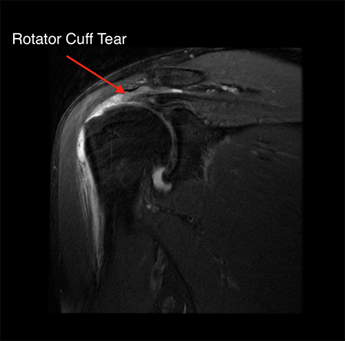
Figure 2. Coronal MRI of the shoulder demonstrating a full-thickness tear of the supraspinatus tendon (arrow). This is a fluid-sensitive sequence, on which fluid appears bright; the bright signal filling the gap where the tendon should attach to the humerus is what identifies the tear.
Two features seen on MRI carry particular weight in decision-making. The first is tendon retraction - how far the torn tendon end has pulled away from its attachment. The second is fatty infiltration, in which the muscle belly is gradually replaced by fat after the tendon tears. Advanced fatty infiltration and muscle atrophy predict both a lower chance of successful repair and poorer results after surgery, and these changes do not reverse once established.22
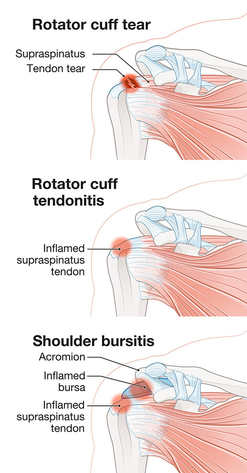
Figure 3. Rotator cuff tear compared with rotator cuff tendonitis and shoulder bursitis. All three can cause pain in a similar location, which is one reason the examination and imaging are used together to establish the diagnosis. (Licensed from Adobe Stock.)
What are the differences between partial and full tears of the rotator cuff?
A full-thickness tear is one in which the tendon is torn all the way through, creating a hole that connects the joint to the space above it. A partial-thickness tear involves only part of the tendon's thickness, so some fibers remain attached.
Partial tears are described by which surface of the tendon is involved. An articular-sided tear involves the undersurface of the tendon, facing the joint. A bursal-sided tear involves the top surface, facing the bursa. An intrasubstance tear occurs within the middle of the tendon with both surfaces intact.23 Articular-sided tears are the most common. A specific and frequently encountered pattern is the PASTA lesion - partial articular-sided supraspinatus tendon avulsion - in which the undersurface of the supraspinatus has pulled away from its attachment on the bone.24
Partial tears are also graded by depth. The Ellman classification is the most widely used: grade I involves less than 25% of the tendon thickness (under 3 mm), grade II involves 25% to 50% (3 to 6 mm), and grade III involves more than 50% (over 6 mm).25 This grading matters because it drives treatment. Low-grade tears are generally managed without repair, while high-grade tears are more often repaired.
Full-thickness tears are described by size: small (less than 1 cm), medium (1 to 3 cm), large (3 to 5 cm) and massive (greater than 5 cm, or involving two or more tendons).8
The practical difference between partial and full tears lies in their natural history. Both types of tear can enlarge over time, but they do so at different rates. In prospective studies following patients with ultrasound and MRI, asymptomatic full-thickness tears enlarged in approximately 22% of shoulders at two years and 50% at five years. Partial-thickness tears progressed more slowly, with enlargement rates of about 11% at two years and 35% at five years.26 Among high-grade partial tears specifically, conversion to a full-thickness tear has been reported in a substantial minority of patients over time.27
Importantly, tears do not spontaneously heal. A torn rotator cuff tendon will not reattach itself. The question is not whether the tear will repair itself but whether it will enlarge, and whether it is causing enough symptoms to warrant treatment.
Tear enlargement matters because it is closely linked to two other things: the development of pain, and the progression of fatty degeneration in the muscle.26 In one prospective cohort, one-third of tears increased by more than 10 mm, and progression of degenerative muscle changes was seen in half of subjects. Shoulder function tended to remain stable if the tear enlarged by less than 20 mm.28
What are the treatment options for a rotator cuff tear?
Treatment is always individualized. The factors I consider include your age, your activity level and demands, whether the tear was traumatic or degenerative, how long you have had symptoms, the size and location of the tear, the degree of retraction and fatty infiltration of the muscle, the condition of the rest of the shoulder, and your general health and healing capacity.
Nonoperative treatment
Nonoperative treatment is a reasonable and well-accepted starting point for most degenerative tears. The core of nonoperative treatment is a structured physical therapy program. The goals are to restore motion, to strengthen the remaining intact rotator cuff and the muscles around the shoulder blade so they can compensate for the torn tendon, and to correct the mechanics of how the shoulder and shoulder blade move together.
This works reasonably well for many patients. In a prospective cohort of patients with rotator cuff-related shoulder pain who completed a standardized progressive exercise program, average night pain fell from 4.3 to 1.6 on a 10-point scale and pain with activity fell from 5.7 to 2.5 over 26 weeks, with significant improvement in motion, strength and quality of life.29 Data from the Rotator Cuff Outcomes Workgroup (ROW) cohort likewise support physical therapy in the nonoperative management of these tears.30
Other nonoperative measures include activity modification, anti-inflammatory medication and corticosteroid injection. A subacromial corticosteroid injection can provide meaningful short-term relief of pain and is often useful both diagnostically and therapeutically.31 The relief is generally temporary, and repeated injections are used judiciously.
It is worth being aware that nonoperative treatment and surgery differ in how quickly they work. In a comparative study of time to improvement, patients treated nonoperatively and patients treated surgically both improved, but along different timelines.32 Nonoperative treatment tends to improve symptoms sooner, while surgery is directed at the underlying structural problem.
The important limitation of nonoperative treatment is that it does not address the tear. In a randomized trial comparing tendon repair with physiotherapy for small-to-medium-sized full-thickness tears, followed out to fifteen years, primary tendon repair remained superior to physiotherapy: an 11.8-point advantage on the Constant score, a 13.9-point advantage on the ASES score, and 1.8 cm less pain on a 10-cm visual analog scale. Among the 26 tears treated with physiotherapy alone, mean tear size increased from 16.2 mm to 31.6 mm. Fifteen of the 51 patients originally assigned to physiotherapy ultimately crossed over to surgery.33
Surgical treatment
Surgical treatment means repairing the torn tendon back to its attachment on the bone. In my practice this is done arthroscopically, through several small incisions, using a camera and instruments placed into the shoulder.
Surgery is generally considered when a reasonable course of nonoperative treatment has not relieved your symptoms, or when the characteristics of the tear favor early repair. Certain tears warrant strong consideration for early surgical repair rather than a prolonged nonoperative trial: acute traumatic full-thickness tears, acute-on-chronic tears with new pseudoparalysis or profound external rotation weakness, and tears in patients whose muscle quality and age remain compatible with healing.28
There is genuine debate in the literature about the benefit of surgery for small degenerative tears. A Cochrane systematic review of three randomized trials comparing repair with nonoperative treatment found that at one year, surgery probably provides little or no clinically important improvement in pain or function compared with exercise therapy. The authors were careful to note that the trials included mostly small degenerative supraspinatus tears, contained methodological concerns, and that their conclusions may not apply to traumatic tears, large tears involving the subscapularis, or younger patients.34
Against this, the fifteen-year randomized data described above favor repair, and prospective longitudinal data show that patients treated surgically outperformed nonoperatively managed patients in nearly all clinical parameters at final follow-up, with a lower prevalence of fatty degeneration in the supraspinatus and infraspinatus.26,33
Surgery is also considered for massive and irreparable tears, though the options there are different and include partial repair, superior capsular reconstruction, tendon transfer, subacromial balloon spacer and, in the setting of arthritis, reverse shoulder replacement.18,35,36 These are beyond the scope of this guide and would be discussed individually if they apply to you.
How is a rotator cuff repair performed?
All of the repairs described below are performed arthroscopically, under a general anesthetic, usually combined with a regional nerve block. Several small incisions, called portals, are made around the shoulder. A camera is placed through one portal and instruments through the others. The joint is inspected first, then the space above the cuff. Any inflamed bursa is removed so the tendon can be seen clearly, the edges of the tear are prepared, and the bone at the attachment site is lightly abraded to encourage healing.
Repairs are held with suture anchors - small implants placed into the bone that carry strong sutures. Modern constructs commonly use soft, all-suture anchors, which are smaller, occupy less of the tendon footprint on the bone, and are easier to revise if a second surgery is ever needed.37 Suture tape is frequently used in place of conventional round suture; the broader profile distributes force over a wider area of tendon and has been associated with lower retear rates in meta-analysis.38
Arthroscopic PASTA repair
A PASTA lesion is a partial tear of the undersurface of the supraspinatus, where the deep fibers have pulled off the bone but the top surface of the tendon remains intact. The surgical question is how to reattach the torn deep fibers without destroying the healthy tissue that remains.
There are two general approaches. In a tear completion repair, the remaining intact fibers are cut through, converting the partial tear into a full-thickness tear, which is then repaired in standard fashion. In an in situ transtendon repair, the intact bursal-sided fibers are preserved, and sutures are passed through the substance of the tendon to bring the torn deep layer back down onto the bone. Systematic review has found that the two techniques yield similar postoperative outcomes, and that the in situ transtendon approach is particularly well suited to PASTA lesions.25
The technique I use is a knotless PASTA bridge repair. Anchors are placed into the footprint on the humerus beneath the intact tendon. Suture from these anchors is passed through the torn undersurface fibers and then brought over the top of the repair site in a bridging pattern, secured laterally with additional anchors. The result is a construct that compresses the torn deep layer of tendon back against its bony footprint over a broad area, without knots sitting on the surface of the tendon and without sacrificing the intact bursal-sided fibers.
In general, partial tears involving less than 50% of the tendon thickness are treated with debridement rather than repair, often with concomitant subacromial decompression, while higher-grade tears are repaired.25 Systematic review of arthroscopic repair of partial-thickness tears has shown improved outcomes with repair.39
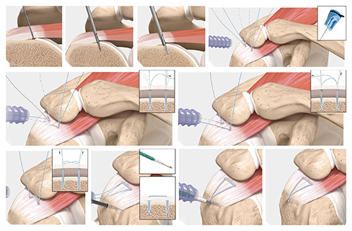
Figure 4. Arthroscopic repair of a PASTA lesion (partial articular-sided supraspinatus tendon avulsion) using a knotless bridging construct. The torn undersurface fibers are secured back to the footprint on the humerus without taking down the intact bursal-sided tendon. (Credit: Arthrex, Inc.)
Arthroscopic knotless double-row repair
For most full-thickness tears I use a knotless double-row, or transosseous-equivalent, repair. Two anchors are placed at the medial edge of the tendon footprint, near the cartilage of the humeral head. Suture tape from these anchors is passed through the tendon. The tapes are then brought laterally, crossed over the top of the tendon, and secured into the outer edge of the greater tuberosity with a second row of anchors.
The purpose of this construct is to press the tendon flat against a wide area of prepared bone rather than fixing it at a single line of points. Cadaveric biomechanical work confirms that double-row and transosseous-equivalent constructs produce greater footprint contact area and higher load to failure than single-row repairs.40
Whether that biomechanical advantage translates into better healing in patients is less clear. In a large meta-analysis, average retear rate was 14.5% for single-row repair and 12.7% for double-row repair, a difference that did not reach statistical significance.41 Systematic review and meta-analysis comparing single- and double-row repair likewise found functional outcomes broadly comparable.42 I favor the double-row construct for the footprint restoration and mechanical stability it provides, particularly in medium and larger tears, while acknowledging that the published difference in healing rates is modest.
The term knotless refers to the absence of tied suture knots over the tendon. Knotless constructs avoid a bulky knot stack sitting under the acromion.
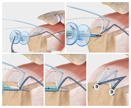
Figure 5. Knotless double-row (transosseous-equivalent) repair of a full-thickness rotator cuff tear. Suture tape from medial anchors is passed through the tendon and then bridged over the tendon to lateral anchors, compressing the tendon against its footprint on the humerus over a broad surface area. (Credit: Arthrex, Inc.)
The Regeneten bioinductive implant
The Regeneten implant (Smith+Nephew) is a small patch made of highly purified bovine collagen. It is placed arthroscopically onto the top surface of the rotator cuff tendon, either over a completed repair or, in some partial tears, on its own without a formal repair. It is held with small staple-like anchors.
The implant is described as bioinductive rather than structural. It does not provide mechanical strength to the repair. Instead, it acts as a scaffold that induces the body to lay down new tendon-like tissue, thickening the existing tendon over the following months. The implant itself is resorbed.
In a systematic review and meta-analysis of thirteen studies, patients treated with the bioinductive patch showed significant improvement in ASES scores, Constant-Murley scores and pain, with most patients achieving the minimal clinically important difference, and significant increases in tendon thickness. The overall retear rate after full-thickness repair augmented with the patch was 8.3%, and after partial-thickness repair 1.1% - lower than historical rates for traditional repair. The overall complication rate was 15.5% for full-thickness and 16.2% for partial-thickness repairs, similar to standard repair for full-thickness tears and somewhat higher for partial-thickness tears. Adverse reactions to the implant itself were reported at 0.2%.44
The authors of that review were appropriately cautious, noting a lack of case-control studies directly comparing the bioinductive patch with traditional repair. My view is that it is a safe addition that appears to reduce retear risk, and I consider it selectively - most often in revision settings, in tears where tissue quality is poor, and in patients with risk factors for failed healing.45
What are other possible procedures at the time of the rotator cuff surgery?
It is common to find and treat other sources of shoulder pain at the same operation. These are usually discussed with you before surgery, but sometimes the decision is made based on what is found once the camera is in the shoulder.
Subpectoral biceps tenodesis
The long head of the biceps tendon runs from the top of the shoulder socket, through the joint, and down a groove in the front of the humerus. It is a well-recognized source of anterior shoulder pain. The tendon is densely supplied with free nerve endings, which supports its role as a pain generator, and biceps pathology very frequently accompanies rotator cuff tears.46
A tenodesis detaches the tendon from its origin inside the joint and reattaches it lower down, outside the joint, to the humerus. This removes the painful intra-articular portion of the tendon while preserving the muscle's length, contour and function. In a subpectoral tenodesis, this reattachment is performed through a small incision at the front of the arm just below the pectoralis major muscle, which places the fixation below the groove and removes the entire length of tendon that can be a source of pain.
The alternative is a tenotomy, in which the tendon is simply released and not reattached. Tenotomy is quicker and is often chosen in older, lower-demand patients. It carries a higher rate of cosmetic deformity - the so-called Popeye deformity, where the muscle belly balls up in the lower arm - and can cause cramping. Contemporary literature suggests functional outcomes are broadly equivalent between the two, aside from the higher rate of cosmetic deformity with tenotomy, and many surgeons opt for tenodesis in younger or higher-demand patients and tenotomy in older, lower-demand patients.46
Arthroscopic distal clavicle excision
The acromioclavicular joint sits at the top of the shoulder, where the collarbone meets the acromion. It is a small joint that commonly develops arthritis, and when it is arthritic it causes pain localized to the top of the shoulder, often worse with reaching across the body.
A distal clavicle excision, historically called a Mumford procedure, removes a small amount of bone - typically about a centimeter - from the outer end of the collarbone. This eliminates contact between the two arthritic surfaces. Performed arthroscopically, it is done through the same portals as the rotator cuff repair.47 It is performed in the presence of painful acromioclavicular joint arthritis, and moderate strength evidence supports its use as a concomitant treatment alongside arthroscopic repair in patients with full-thickness rotator cuff tears and symptomatic acromioclavicular arthritis.47
Arthroscopic subacromial decompression
Subacromial decompression, or acromioplasty, involves shaving the undersurface of the acromion with a burr to increase the space above the rotator cuff, along with removing inflamed bursa. The rationale is to reduce mechanical contact between the bone above and the tendon below.
The role of acromioplasty performed alongside rotator cuff repair has been studied extensively, and the evidence is genuinely mixed. Several meta-analyses have found that repairs with and without acromioplasty yield similar clinical outcomes.48 A recent analysis found that the functional outcome results in this literature are statistically fragile, meaning small changes in a handful of patients would reverse the published conclusions.49
However, the long-term follow-up of a multicenter randomized controlled trial is informative. At a mean of 11 years, there was no difference in patient-reported outcome scores between patients who did and did not have an acromioplasty. But 7 of 45 patients (16%) in the group without acromioplasty underwent reoperation, compared with 1 of 41 patients (2%) in the acromioplasty group. Every patient who required reoperation had a Type 2 or Type 3 (curved or hooked) acromion.50 A systematic review analyzing outcomes by acromial type reached a similar conclusion.51
My approach is therefore selective. I perform an acromioplasty when the acromial shape and the appearance of the undersurface at the time of surgery suggest that mechanical contact is contributing to the problem, and I do not perform it routinely in patients with a flat acromion.
When should I have my rotator cuff repaired?
The answer differs substantially depending on whether your tear is traumatic or degenerative.
For acute traumatic full-thickness tears, earlier repair is preferable. A systematic review and meta-analysis of thirteen studies comprising 871 patients found that early repair was associated with a significantly lower retear rate than delayed repair (risk ratio 0.60), and four studies demonstrated significantly greater functional improvement in the early intervention group. No study found early intervention to be detrimental or delayed intervention to be superior.52 Traumatic tears also carry a real risk of retraction and muscle degeneration if left, which can make a technically straightforward repair much more difficult later.
That said, delay is not always catastrophic. A study specifically examining traumatic tears found that delayed surgery did not necessarily worsen functional outcomes,53 and a series of traumatic full-thickness tears initially treated nonoperatively found that a portion of patients did reasonably well without surgery, although satisfaction was higher in those who ultimately had surgery (83% versus 55%).54
For atraumatic degenerative tears, the timeline is less pressing. In a retrospective cohort comparing patients who had surgery within 12 months of symptom onset with those who had surgery after at least 12 months, there was no difference in retear rate (12% versus 9%), reoperation rate (5% versus 3%), postoperative range of motion, strength or patient-reported outcome scores.55 This means there is generally time to give nonoperative treatment a fair trial.
What does drive the timing in degenerative tears is not the calendar but the biology. The features that make a repair likely to succeed - limited retraction, preserved muscle quality without advanced fatty infiltration, and an age compatible with healing - can deteriorate over time. Median time from a tear enlargement event to progression of muscle degeneration has been measured at approximately one year for the supraspinatus and infraspinatus.28
Ten-year data from the MOON Shoulder cohort found that the predictors of eventually undergoing surgery for a symptomatic atraumatic tear change over time, which reinforces the value of ongoing follow-up rather than a single decision made at one point.56
What are the outcomes of rotator cuff repair surgery?
Most patients do well. Rotator cuff repair reliably reduces pain and improves function, and patient satisfaction is high. Improvements in pain scores are typically large: in one systematic review of patients over 70 years of age, mean pain score fell from 6.3 cm to 1.7 cm on a visual analog scale, a difference of 4.6 cm - nearly three times the minimal clinically important difference.57
Range of motion also improves. A systematic review of range of motion following arthroscopic repair documented significant gains in forward flexion, abduction and rotation across the published literature.58
Healing of the repair, however, is not universal. This is the single most important thing to understand about rotator cuff surgery. Retear rates reported in the literature range widely - from 11% to more than 90% - depending on patient characteristics, tear characteristics, tissue quality and surgical technique.59 Pooled retear rates in large meta-analyses fall in the 15% to 20% range overall.41
The two strongest predictors of failed healing are age and tear size. In pooled analysis, patients under 60 years of age had a retear rate of 14.4%, compared with 24.3% for patients over 60. Small and medium tears had an average retear rate of 12.5%, compared with 37% for large and massive tears.41 Data from a large randomized controlled trial similarly demonstrated that increasing age and increasing tear size independently reduce healing rate at one year.60
A crucial and somewhat counterintuitive point: a retear is not the same as a failed operation. Many patients whose repair does not heal completely still experience substantial and durable pain relief and functional improvement. That said, patients with intact repairs do score better on average. In a prospective cohort study correlating functional outcome scores with MRI-based structural integrity, patients with intact repairs had significantly better scores than patients with failed repairs.61
Table 1. Retear rates after arthroscopic rotator cuff repair, by patient and repair factor.
|
Factor |
Retear rate |
Comment |
|
Age under 60 years |
14.4% |
Younger age favors healing (OR 1.8 for older age) |
|
Age 60 years and over |
24.3% |
|
|
Small to medium tears |
12.5% |
Larger tears strongly predict failure (OR 0.3) |
|
Large to massive tears |
37% |
|
|
Single-row repair |
14.5% |
No statistically significant difference between constructs |
|
Double-row repair |
12.7% |
|
|
Repair with tendon augmentation |
21.2% |
Augmentation used in higher-risk tears |
|
Repair without augmentation |
51.2% |
|
|
Repair with PRP |
14.5% |
Lower retear rate with PRP in pooled analysis |
|
Repair without PRP |
23.9% |
|
|
Revision repair |
25.3% |
From a separate systematic review |
Data from Longo et al, BMC Musculoskelet Disord. 2021;22:749, except revision data from Hurley et al, Am J Sports Med. 2023;51:3080-3087.
Several factors that influence healing are within your control. Smoking has a clear detrimental effect on both clinical and structural outcomes after repair.62 This includes heated tobacco products, which have been shown to impair rotator cuff healing to a degree similar to conventional cigarettes.63 Nutritional status also matters: preoperative nutrition has been shown to impact retear rate after arthroscopic repair.64 Control of diabetes is likewise important.65
If a repair does fail and symptoms warrant further surgery, revision repair is possible. A systematic review of revision arthroscopic repair found moderate functional outcomes with a retear rate of 25.3%, with mean ASES score of 75.0 and satisfaction of 78.4%.66
When can I return to sports and work after rotator cuff surgery?
Return to work depends far more on what your work requires than on the surgery itself.
In a systematic review and meta-analysis, between 59.5% and 97% of patients returned to work, at an average of 5 to 9 months postoperatively. Heavy manual labor is a risk factor for not returning to a prior level of work.67 In a large single-surgeon series of 1,502 primary arthroscopic repairs, 76% of patients had returned to work at six months, and 40% had returned to their preinjury level of work.68
Patients with physically demanding work have significantly worse time to return to work, lower levels of employment on return, and higher rates of job loss than patients in sedentary occupations.69 Among manual laborers specifically, one series found 89.6% were able to maintain manual labor positions and had returned to work at an average of approximately 8 months.70 Workers' compensation status has been associated with poorer return-to-work outcomes in several studies, though this is not a universal finding.71
If your work is sedentary, you may be able to return within a few weeks, working within the restrictions of the sling. If your work involves overhead activity or lifting, expect a longer absence, and light duty is very helpful if your employer can accommodate it.
For sport, a systematic review of overhead athletes undergoing arthroscopic repair found that 75.4% returned to play at a mean of 6.4 months, and 62.5% returned to sport at their preinjury level. The overall complication rate in that population was 7.1%, and 10.1% underwent a further reoperation.72 Return to sport in athletes older than 35 years has been shown to be achievable.73 In general, return to sport and recreational activity is not permitted sooner than four to six months, and always with clearance from both surgeon and therapist.
Table 2. Return to work and return to sport after arthroscopic rotator cuff repair.
|
Outcome |
Rate |
Timing |
|
Return to work (any level), pooled |
59.5% - 97% |
Mean 5 - 9 months |
|
Return to work (any level) at 6 months |
76% |
6 months |
|
Return to preinjury level of work at 6 months |
40% |
6 months |
|
Return to manual labor |
89.6% |
Mean ~8 months |
|
Return to play, overhead athletes |
75.4% |
Mean 6.4 months |
|
Return to preinjury level of sport, overhead athletes |
62.5% |
- |
|
Reoperation, overhead athletes |
10.1% |
- |
Data compiled from Khan et al, Clin Sports Med. 2023;42:125-140; Ting et al, Am J Sports Med. 2023;51:923-932; Green et al, Am J Sports Med. 2022;50:2227-2233; Migliorini et al, J Orthop Traumatol. 2023;24:3.
What are the possible complications of rotator cuff repair surgery?
Arthroscopic rotator cuff repair is a safe operation, and serious complications are uncommon. Observational data from surgical registries indicate that the risk of serious adverse events such as deep infection, pulmonary embolism or death is likely less than 1%.34 Nonetheless, complications do occur and you should understand them before proceeding.
Retear or failure of healing
This is the most common complication and is discussed in detail above. Most recurrent tears occur within the first six months after surgery.74 Risk factors include older age, larger tear size, fatty infiltration and atrophy of the muscle, poorer tissue quality, smoking, diabetes, and poor bone quality - one study reported retear rates of 9% in patients with normal bone mineral density, 30.2% in patients with osteopenia and 41.7% in patients with osteoporosis.65
Postoperative shoulder stiffness
Stiffness is the second most common problem. In a systematic review and meta-analysis of 18 studies including 63,565 patients, the identified risk factors for postoperative stiffness were advanced age, female sex (odds ratio 1.99), diabetes (odds ratio 2.01), thyroid disease (odds ratio 1.32) and workers' compensation insurance (odds ratio 1.92).75 Most stiffness resolves with time and therapy. Occasionally a manipulation under anesthesia or arthroscopic release is required.
Infection
Infection after arthroscopic shoulder surgery is uncommon, with rates generally well below those of open surgery. The shoulder is unusual in that the most common organism is Cutibacterium acnes, a slow-growing bacterium that lives in the hair follicles and sebaceous glands of the skin around the shoulder and can present in a delayed and subtle fashion. Prevention includes sterile skin preparation and antibiotics at the time of surgery.76 Signs of infection include fever, chills, increasing pain and swelling, redness, warmth and drainage. Treatment may require antibiotics and possibly a return to the operating room for irrigation and debridement.
Nerve injury
Major nerve injury is rare. Temporary numbness or altered sensation around the portals is more common and usually resolves. Nerve blocks used for anesthesia carry their own small risk of temporary nerve symptoms; long-term deficits are uncommon.77 Complications specific to the interscalene block include temporary hoarseness, Horner syndrome and temporary diaphragm weakness from phrenic nerve blockade.77
Persistent pain
Some patients continue to have pain despite an intact repair. This may relate to the biceps tendon, the acromioclavicular joint, cervical spine pathology, or glenohumeral arthritis. Careful preoperative evaluation reduces but does not eliminate this possibility.
Other reported complications
Other complications reported after arthroscopic rotator cuff repair include anchor pullout or migration, damage to the articular cartilage from instruments or anchors, tendon lengthening at the repair site with resulting weakness,78 deep venous thrombosis, complications of anesthesia, and complex regional pain syndrome. In a series of manual laborers, the overall postoperative complication rate was 8.33%.70
This is not an exhaustive list of all complications. Other complications, although uncommon, can still occur.
Table 3. Reported rates of the more common complications after arthroscopic rotator cuff repair.
|
Complication |
Reported rate |
Principal risk factors |
|
Retear / failure of healing (overall) |
~15%-20% pooled; 11%-90% range |
Age, tear size, fatty infiltration, smoking, diabetes, poor bone quality |
|
Retear after revision repair |
25.3% |
Prior failed repair |
|
Postoperative shoulder stiffness |
Variable by series |
Age, female sex (OR 1.99), diabetes (OR 2.01), thyroid disease (OR 1.32), workers' compensation (OR 1.92) |
|
Serious adverse events (deep infection, PE, death) |
<1% |
General health, comorbidity |
|
Overall complication rate, manual laborers |
8.33% |
Occupational demand |
|
Overall complication rate, overhead athletes |
7.1% |
- |
|
Reoperation, overhead athletes |
10.1% |
- |
|
Reoperation after repair without acromioplasty (11-yr) |
16% |
Type 2 or 3 acromion |
|
Reoperation after repair with acromioplasty (11-yr) |
2% |
- |
Data compiled from Longo et al, BMC Musculoskelet Disord. 2021;22:749; Hurley et al, Am J Sports Med. 2023;51:3080-3087; Wang et al, JBJS Rev. 2025;13(10):e25.00134; Karjalainen et al, Cochrane Database Syst Rev. 2019;12:CD013502; Green et al, Am J Sports Med. 2022;50:2227-2233; Migliorini et al, J Orthop Traumatol. 2023;24:3; Woodmass et al, J Bone Joint Surg Am. 2022;104:2101-2107.
What will help with my pain after surgery?
Arthroscopic rotator cuff repair is associated with significant postoperative pain, with 30% to 70% of patients reporting significant pain following surgery. Pain is a primary obstacle to starting physical therapy, and delayed therapy is associated with poorer functional outcomes, so controlling it well matters for more than comfort alone.79
Pain control after your surgery uses several methods together rather than relying on narcotics alone. This is called a multimodal approach. A prospective randomized controlled trial found that a multimodal nonopioid protocol provided better or equivalent pain control compared with opioid analgesia following arthroscopic rotator cuff surgery.80 When opioids are prescribed, they should be taken at the lowest effective dose for the shortest period.77
Frequent icing of the shoulder, three to four times per day, is a simple and effective adjunct.
Single-shot nerve block
Most patients receive a single-shot interscalene brachial plexus nerve block before surgery. Local anesthetic is injected around the nerves in the side of the neck that supply the shoulder and arm, under ultrasound guidance. This numbs the shoulder for the operation and for a period afterward.
A network meta-analysis of 14 randomized controlled trials including 851 patients found that peripheral nerve blocks are highly effective at attenuating postoperative pain after arthroscopic rotator cuff repair and should be more widely considered as an alternative to general anesthesia alone. Single-shot interscalene block was the most commonly used technique, accounting for 37.8% of patients in the pooled data.79
There are two practical things to know. The first is that your arm will be numb and weak - often completely - for a period after surgery. This is expected and temporary. Typically the block wears off between 8 and 24 hours after it is placed. The second, and more important, is that you should start your pain medication before the block wears off. If you wait until the block has worn off and the pain is severe, it is much harder to get ahead of it.
Effective pain management after arthroscopic repair has been shown to reduce length of stay by approximately 9.6%.79 Reported complications specific to the interscalene block include temporary hoarseness, Horner syndrome and phrenic nerve palsy causing temporary weakness of the diaphragm on that side.77
Will platelet rich plasma help with healing of my rotator cuff repair surgery?
Platelet-rich plasma (PRP) is produced by drawing your own blood and spinning it in a centrifuge to concentrate the platelets. Platelets release growth factors, and the rationale is that delivering these growth factors to the repair site may reduce inflammation and support tendon and soft tissue healing.45
There is some evidence in favor. In pooled analysis of retear rates, the average retear rate was 14.5% in repairs performed with PRP compared with 23.9% in repairs performed without it, a statistically significant difference.41 Systematic reviews have demonstrated that intraoperative PRP injections improve pain and function in the short term for both full-thickness and partial rotator cuff tears, with reduced retear rates in full-thickness tears.45 A prospective randomized in vivo study examining the biological effect of PRP on rotator cuff tears has also been performed.82
Do I need to use a sling after rotator cuff surgery?
Yes. You will be placed in a sling, usually with a small foam abduction pillow between your arm and your body, immediately after surgery.
The sling protects the repair during the period when the tendon is healing to bone. This healing is biological and takes time; the repair is at its most vulnerable in the early weeks. You will wear the sling for four to eight weeks depending on the size of your tear and the extent of the repair, and it should be worn at all times, including at night.
The foam cushion should be left in place at all times while wearing the sling. While in the sling, the elbow should be bent at a right angle and the hand should be level with the elbow or slightly higher, with the elbow resting slightly in front of your body.
You may remove the sling for the specific exercises prescribed by your surgeon or therapist, for icing, for dressing and for showering. Outside of those activities it stays on.
The duration of immobilization is a subject of ongoing study, and the evidence does not clearly favor one approach.
When can I drive after rotator cuff surgery?
There is no definitive test to determine when a patient is safe to return to driving, and recommendations have historically been conservative.
That said, several practical conditions must be met before you drive:
- You must not be taking narcotic or other sedating medications.
- You must be able to drive without wearing the sling, and you must be comfortable and safe doing so.
- You must have sufficient strength and range of motion to control the steering wheel in all situations, including an emergency maneuver - not merely in normal straight-line driving.
- You should be confident, not merely willing. If you are uncertain, practice in an empty parking lot before driving in traffic.
What does rehabilitation look like after rotator cuff surgery?
Rehabilitation after rotator cuff repair is staged, and the stages exist for a biological reason. The repaired tendon must heal to bone, and that healing determines how quickly load can be applied. Rehabilitation is individualized based on repair integrity, tissue quality, and patient factors such as revision status and smoking history.87
The program is generally described in four stages:87
- Stage 1 focuses on allowing anatomic rotator cuff healing while maintaining passive range of motion.
- Stage 2 aims to protect the repair while reestablishing dynamic shoulder stability and developing full active and passive range of motion.
- Stage 3 maintains shoulder range of motion while progressing with shoulder stabilization exercises and adding functional shoulder exercises.
- Stage 4 restores full functional strength and implements sport-specific training if necessary.
Formal physical therapy can usually be initiated within the first two to six weeks following surgery, and I will refer you when the timing is appropriate. This includes sessions at the clinic in addition to a home exercise program for range of motion and postural exercises. For very large tears, physical therapy may be delayed to promote rotator cuff healing, up to six weeks postoperatively.
The timing of therapy has been studied. A study examining the effect of physical therapy timing on rotator cuff repair revisions and capsulitis found relationships between when therapy begins and both of these outcomes.88 Pooled data suggest that beginning active-assisted range of motion before five weeks corresponds to a higher retear rate than beginning after five weeks (25.6% versus 14.2%), while beginning full active range of motion before eight weeks corresponds to a lower retear rate than beginning after eight weeks (12.1% versus 21.8%).41 In other words, the early weeks are the ones that require genuine protection, but once that window has passed, delay carries its own cost. Slower rehabilitation postoperatively has not been shown to increase rates of stiffness at one year.87
Two tear-size-specific protocols are used in my practice - one for small-to-medium repairs and one for large-to-massive repairs - and the appropriate one will be provided to you and to your therapist.
The two protocols below are the ones used in my practice. Which one applies to you depends on the size of the tear that was repaired, and I will tell you which you are following. Bring the protocol to your first physical therapy visit. Timelines are guidelines rather than deadlines; your therapist and I will adjust them based on how your shoulder responds.
Table 4. Rehabilitation protocol after arthroscopic rotator cuff repair - small and medium tears.
|
Phase |
Goals |
Exercises and milestones |
|
Phase 1 Weeks 1-6 Sling × 6 weeks (sleep included) |
1. Minimize pain and inflammation. 2. Achieve staged ROM goals (avoid aggressive PROM). 3. Protect the repair. 4. Scapular stabilization. 5. Discontinue abduction pillow at 4 weeks; sling for 2 weeks following. 6. No active shoulder ROM, lifting, supporting body weight, or lifting body weight with the hands. |
Days 1-10: Pendulum hangs (no active movement). Finger, wrist and elbow AROM (no weight) - elbow PROM only if a biceps tenodesis was performed. Seated scapular isometrics and cervical ROM. Begin PROM flexion 60-90° and ER at 20° abduction 0-15°, in the plane of the scapula. Weeks 2-3: Continue PROM progression - flexion 60-100°, ER at 20° abduction 0-20°. No internal rotation or extension PROM. Begin resisted finger, wrist and elbow AROM. Resume general conditioning (walking, stationary bicycle; no treadmill walking or elliptical). Begin manual scapular strengthening. Weeks 4-6: Continue PROM progression - flexion 90-125°, ER at 20° abduction 15-40°. For subscapularis repairs, ER at 0° abduction 0-30° PROM/AAROM for 6 weeks unless otherwise directed. Begin joint mobilizations (grades 1 and 2) as tolerated. Progress scapular isometrics (sidelying retractions). For biceps tenodesis, delay elbow flexion strengthening 6 weeks. Avoid UBE. Criteria to progress: staged ROM goals achieved, minimal to no pain. |
|
Phase 2 Weeks 7-12 |
1. Allow soft-tissue healing; do not over-stress the repair. 2. Gradually restore full PROM by week 12. 3. Minimize pain and inflammation. Restrictions out of the sling: no lifting anything heavier than a coffee cup; no ROM beyond staged goals or excessive behind-the-back movement; no supporting body weight with the hands or arms; no sudden jerking motions; no long-lever rotator cuff strengthening that can stress the repair; no empty-can exercises at any stage. |
Begin gentle scapular and glenohumeral mobilizations to regain full PROM. Progress from prone scapular retractions to prone rows to neutral extensions. Begin PROM in other planes (horizontal adduction; ER at 45°, 70° and 90° abduction). Week 7: PROM flexion 120-140°, ER at 20° abduction 30-60°, ER at 90° abduction 40-60°. AAROM as tolerated. Week 9: PROM flexion 130-155°, ER at 20° abduction 45-60°+, ER at 90° abduction 50-75°, AROM flexion 80-120°. Weeks 10-12: AROM with focus on good mechanics - sidelying to supine to standing scaption; supine to sidelying ER with a towel under the arm as needed; supine protraction; push-up plus (wall to table to bench to floor); prone rows; prone extension with ER; prone horizontal abduction with ER; prone scaption from week 9. When mechanics are good and motion is pain-free, begin strengthening of the deltoid, non-repaired cuff segments and scapular musculature - light resistive band work in a pain-free range, scapular strengthening, low-level closed-chain program. Neuromuscular re-education for scapular mobility and core stability. Criteria to progress: staged ROM goals achieved with minimal pain, strengthening tolerated with minimal pain, good static and dynamic posture. |
|
Phase 3 Months 3-6 |
1. Full pain-free PROM and AROM. 2. Enhance dynamic shoulder stability and neuromuscular control. 3. Gradual restoration of shoulder strength and endurance. 4. Gradual return to full functional activities. 5. Avoid lifting objects of 15-20 lb, sudden lifting or jerking, and overhead lifting. |
Week 12: PROM flexion 140° to within normal limits, ER at 20° abduction within normal limits, ER at 90° abduction 75° to within normal limits, AROM flexion 115-145°+. Begin functional internal rotation stretch (behind the back). Begin light PNF diagonals for the cuff, deltoid and scapula. Begin open-chain rhythmic stabilizations. Begin closed-chain activity progression. Begin the advanced strengthening program as tolerated - criteria: manual muscle testing at least 4/5, pain-free basic activities of daily living and initial strengthening program, full AROM elevation. Sample exercises: standing band PNF, band 90/90 ER and IR, band sport simulations. Criteria to progress: adequate strength and dynamic stability for higher-demand work or sport-specific activity. |
|
Phase 4 Month 7 and beyond |
1. Maintain full pain-free ROM. 2. Advanced conditioning - muscular strength, power and endurance. 3. Begin return-to-sport training. |
Continue strengthening progression into sport-specific programs (Thrower's 10). Progressive return to a weight-lifting program: begin with light weight and high repetitions, progressing to higher weight and lower repetitions. Begin a plyometric program. Initiate an interval sport program after a successful 3-6 week plyometric program. Criteria before return to work or sport: physician clearance, no pain at rest, minimal pain with activity, no sensation of instability, sufficient ROM for the desired activity, and adequate strength and endurance of the rotator cuff and scapular musculature with minimal to no pain or difficulty. |
Table 5. Rehabilitation protocol after arthroscopic rotator cuff repair - large and massive tears.
|
Phase |
Goals |
Exercises and milestones |
|
Phase 1 Weeks 1-7 Sling × 8 weeks (sleep included) Avoid internal rotation and extension |
1. Minimize pain and inflammation. 2. Protect the repair and allow healing. |
Pendulums (no active movement) and shoulder shrugs. Elbow, wrist and hand AROM; ball squeezes. PROM forward elevation in the scapular plane (supine, to 90°) at physical therapy, one visit per week. |
|
Phase 2 Weeks 8-12 |
Restore passive motion and begin active motion while continuing to protect the repair. |
Discontinue the sling. Progress PROM to full in all planes. Achieve full PROM in ER at 20° and at 90° abduction. Begin posterior capsule stretching. Submaximal isometrics with the elbow flexed to 90°. Theraband scapular retractions and periscapular strengthening (low weight, high repetitions). Begin AAROM flexion and progress to AROM flexion. |
|
Phase 3 Months 3-4 |
1. Achieve staged ROM goals. 2. Minimize pain. 3. Improve strength, endurance and power. 4. Increase functional activities. |
Full ROM in all planes. Progress isometrics and the periscapular strengthening program. Closed-chain exercises for dynamic scapular, deltoid and cuff stability. Begin light PNF D1 and D2 patterns and manual resistance for the cuff, deltoid and scapula. Begin Theraband IR and ER strengthening and progressive serratus anterior strengthening. Progress to isotonic dumbbell exercises for the deltoid and supraspinatus (3 lb maximum). Strengthening should emphasize high repetitions and low weight, no more than twice daily. |
|
Phase 4 Months 5-6 |
1. Normalize strength, endurance and power. 2. Return to full activities of daily living and recreational activities. |
Stretching as needed. Continue deltoid, cuff and scapular strengthening (5 lb maximum for isotonic work), progressing by: prone isotonic strengthening; decreasing external stabilization provided to the shoulder girdle; integrating functional patterns; increasing speed of movement; integrating kinesthetic awareness exercises; and decreasing rest time to build endurance. Progress closed-chain dynamic stability activities. Begin isokinetic strengthening. |
|
Phase 5 Months 7-8 |
Advanced conditioning and preparation for return to sport. |
Stretching as needed. Initiate a plyometric program if warranted - two-hand tosses, progressing to one-hand stability drills and then one-handed tosses, varying the amount of abduction and using protected external rotation. Criteria to begin: 5/5 manual muscle testing for the cuff and scapula; start with light weight. Continue the deltoid, cuff and scapular strengthening program. Criteria before return to sport: physician clearance, 5/5 manual muscle testing, completion of the plyometric program if warranted, and completion of an interval return-to-sport program if warranted. |
Post-operative instructions
Immediately after surgery
- Following your stay in the recovery room and when your vital signs are stable, you will be discharged to your escort.
- Remember, it is normal to feel a little dizzy or drowsy for several hours after surgery. This is due to the action of the medicine used during surgery.
- If you do not have a post-operative appointment scheduled, please call the office as soon as possible to schedule this appointment.
- Take your pain medicine as directed. Begin the pain medicine before you start getting uncomfortable, as the nerve block will wear off. If you wait to take your pain medication until the pain is severe, you will have more difficulty in controlling the pain.
- If you are taking narcotic pain medication you may need a stool softener to prevent constipation. Over-the-counter medication such as Docusate or Milk of Magnesia is recommended.
- Use ice on the shoulder three to four times per day. This will help decrease swelling and pain.
- Notify the office of any fever, chills, or temperature > 100.5.
- Notify the office of any wound drainage.
Post-surgery diet
Resume your diet as tolerated and include vegetables, fruits, and proteins (such as meats, fish, chicken, nuts, and eggs) to promote healing. Also, remember to have adequate fluid intake. It is common after surgery to lack an appetite. This may be the result of anesthesia and the medications. Proper nutrition is needed for healing. During the healing process, the body needs increased amounts of calories, protein, vitamins A and C, and sometimes the mineral zinc. Eat a variety of foods to get all the calories, proteins, vitamins, and minerals you need. If you have been told to follow a specific diet, please follow it. What you eat can help heal your wounds and prevent infection and potential complications. This is not merely general advice: preoperative nutritional status has been shown to impact retear rate after arthroscopic rotator cuff repair.64 If you are not eating well after surgery, contact your healthcare provider about nutritional supplements.
How should I manage my surgical site and bandages?
- You can remove your surgical dressing on the third day after your surgery.
- Allow the steri-strips to fall off on their own. If necessary, sutures will be removed at your first post-operative visit.
- You may shower with a waterproof bandage after three days, but keep the incisions dry until the sutures are removed at seven to ten days. Try not to let the direct spray of water from the showerhead hit the incision.
- You can cover your incision with a bandage if needed to prevent irritation from clothing.
- A small amount of drainage from the surgical sites is common. If this occurs, you may apply a bandage to the affected area.
- Swelling in the affected extremity from your shoulder to your hand is common.
- Leakage immediately after surgery is normal and helps to drain some of the fluid that accumulates in the joint during surgery. The dressings may become moist or blood-stained; this is normal and usually not a cause for alarm.
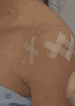
Example of steri-strips.
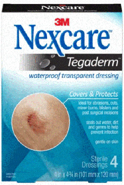
Example of a waterproof bandage.
Do I need to wear a sling or immobilizer?
- You will wear your sling for four to eight weeks depending on the size of your tear and the extent of the repair, and the sling should be worn at all times.
- You may have a small foam cushion between your arm and your body. This should be left in place at all times while wearing the sling.
- While in the sling, the elbow should be bent at a right angle and the hand should be level with your elbow or slightly higher. The elbow should be resting slightly in front of your body.
- You may remove the sling for exercises as prescribed by the surgeon or therapist, for icing, for dressing, and for showering.
What are my precautions regarding movements and positions after surgery?
- Do not actively move your arm away from your body for four to six weeks following surgery. In addition, do not lift any object, even if you are just bending your elbow.
- Follow your surgeon's or therapist's directions regarding rotating your forearm away from your body, to protect the repaired tendon and muscles.
- When lying on your back, we recommend you put a towel roll under your elbow to support the arm.
- Many patients find that lying on a 30-degree incline wedge in bed is more comfortable than lying flat. Some patients find sleeping in a recliner more comfortable during the first few weeks after surgery.
- Initially after surgery you should not reach behind your back with your operated arm.
- Absolutely no lifting.
- You may begin pendulum hangs, but we do not want you to actively move your arm.
- You should start shoulder blade pinches and rolls.
- Passive range of motion for the elbow if you had any surgical work done to your biceps tendon (tenodesis or tenotomy). Active range of motion elbow curls, with no weight, if you did not have any surgical work done to your biceps.
- You can begin active exercises for your wrist and hand, including ball squeezes.
May I use my involved arm for dressing, bathing, driving and other daily activities?
- You may use your wrist, hand and elbow for daily activities. This includes eating, shaving and dressing, as long as you do not move your operated arm away from your body and it does not increase your pain.
- Do not use your arm to push up or off the bed or a chair for at least six weeks after your surgery.
- When using your keyboard and mouse, do not move your arm away from your body.
- When showering, you may wash under the involved armpit by bending forward to let the involved arm hang freely and reaching under with the opposite arm.
- Do not actively move your arm away from your body.
When should I begin formal physical therapy, and how often do I need to go?
- Physical therapy can usually be initiated within the first six weeks following surgery. This includes physical therapy sessions at the clinic in addition to a home exercise program for range of motion and postural exercises. You will eventually be prescribed strengthening exercises.
- For very large tears, physical therapy may be delayed to promote rotator cuff healing, up to six weeks postoperatively.
- Frequency of treatment will be determined by your specific needs together with the physician and physical therapist recommendations.
Can I smoke following surgery?
You should not smoke after surgery, as it interferes with tendon healing. This is not a formality. Systematic review and meta-analysis has shown that smoking has significant detrimental effects on both clinical and structural outcomes after rotator cuff repair.62 Heated tobacco products are not a safe substitute; they have been shown to have detrimental effects on rotator cuff healing similar to conventional cigarettes.63
Exercises you can perform at home at this time
- Pendulum circles. Shift your body weight in circles to allow your operated arm to swing in circles freely. Your operated arm should be fully relaxed.
- Shrugs. Raise your shoulders upward towards your ears. Shrug both shoulders at the same time.
- Scapular retractions. Draw your shoulder blades back and down.
- Elbow active and passive range of motion, and wrist and hand active range of motion.
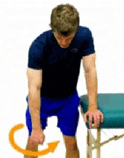
Pendulum circles.
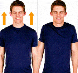
Shrugs.
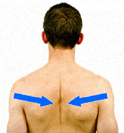
Scapular retractions.
Questions or concerns
If at any time you have questions or concerns, you can either contact your surgeon's medical assistant via email or call the main office at 855-624-3306. You can also use the electronic medical record's online portal to send questions.
References
- Arrillaga B, Miguel-Pérez M, Möller I, et al. Human shoulder anatomy: new ultrasound, anatomical, and microscopic perspectives. Anat Sci Int. 2024;99(3):290-304. doi:10.1007/s12565-024-00775-5
- Yeung A, Fernando A, Patel M, Gatto L, Ackland DC. Muscle and joint function in the rotator cuff deficient shoulder. J Orthop Res. 2024;42(9):2131-2139. doi:10.1002/jor.25909
- Yuri T, Trevino JH 3rd, Hoshikawa K, Hooke A, Giambini H. Moment arms of the anatomical subregions of the rotator cuff muscles during shoulder rotation. Clin Biomech (Bristol). 2023;107:106040. doi:10.1016/j.clinbiomech.2023.106040
- Adams CR, DeMartino AM, Rego G, Denard PJ, Burkhart SS. The rotator cuff and the superior capsule: why we need both. Arthroscopy. 2016;32(12):2628-2637. doi:10.1016/j.arthro.2016.08.011
- Lanham NS, Swindell HW, Levine WN. The subacromial bursa: current concepts review. JBJS Rev. 2021;9(11):e21.00110. doi:10.2106/JBJS.RVW.21.00110
- Johnson J, von Stade D, Gadomski B, et al. Biomechanical and histological changes secondary to aging in the human rotator cuff: a preliminary analysis. J Orthop Res. 2023;41(10):2221-2231. doi:10.1002/jor.25529
- Griffith KM, Hammer LC, Iannuzzi NP, et al. Review of human supraspinatus tendon mechanics. Part I: fatigue damage accumulation and failure. J Shoulder Elbow Surg. 2022;31(12):2671-2677. doi:10.1016/j.jse.2022.06.017
- Dickinson RN, Kuhn JE. Nonoperative treatment of rotator cuff tears. Phys Med Rehabil Clin N Am. 2023;34(2):335-355. doi:10.1016/j.pmr.2022.12.002
- Song A, Cannon D, Kim P, et al. Risk factors for degenerative, symptomatic rotator cuff tears: a case-control study. J Shoulder Elbow Surg. 2022;31(4):806-812. doi:10.1016/j.jse.2021.10.006
- Grusky AZ, Giri A, O'Hanlon D, Jain NB. The relationship of aging and smoking with rotator cuff disease: a systematic review and meta-analysis. Am J Phys Med Rehabil. 2022;101(4):331-340. doi:10.1097/PHM.0000000000001820
- Dabija DI, Gao C, Edwards TL, Kuhn JE, Jain NB. Genetic and familial predisposition to rotator cuff disease: a systematic review. J Shoulder Elbow Surg. 2017;26(6):1103-1112. doi:10.1016/j.jse.2016.11.038
- Giri A, O'Hanlon D, Jain NB. Risk factors for rotator cuff disease: a systematic review and meta-analysis of diabetes, hypertension, and hyperlipidemia. Ann Phys Rehabil Med. 2023;66(1):101631. doi:10.1016/j.rehab.2022.101631
- Kim JH, Min YK, Gwak HC, Kim CW, Lee CR, Lee SJ. Rotator cuff tear incidence association with critical shoulder angle and subacromial osteophytes. J Shoulder Elbow Surg. 2019;28(3):470-475. doi:10.1016/j.jse.2018.08.026
- Moran TE, Werner BC. Surgery and rotator cuff disease: a review of the natural history, indications, and outcomes of nonoperative and operative treatment of rotator cuff tears. Clin Sports Med. 2023;42(1):1-24. doi:10.1016/j.csm.2022.08.001
- Patel M, Amini MH. Management of acute rotator cuff tears. Orthop Clin North Am. 2022;53(1):69-76. doi:10.1016/j.ocl.2021.08.003
- Nyffeler RW, Schenk N, Bissig P. Can a simple fall cause a rotator cuff tear? Literature review and biomechanical considerations. Int Orthop. 2021;45(6):1573-1582. doi:10.1007/s00264-021-05012-6
- Ichinose T, Shitara H, Tajika T, et al. Factors affecting the onset and progression of rotator cuff tears in the general population. Sci Rep. 2021;11(1):1858. doi:10.1038/s41598-020-79867-x
- Shepet KH, Liechti DJ, Kuhn JE. Nonoperative treatment of chronic, massive irreparable rotator cuff tears: a systematic review with synthesis of a standardized rehabilitation protocol. J Shoulder Elbow Surg. 2021;30(6):1431-1444. doi:10.1016/j.jse.2020.11.002
- American Academy of Orthopaedic Surgeons. Management of Rotator Cuff Injuries: Evidence-Based Clinical Practice Guideline. Rosemont, IL: American Academy of Orthopaedic Surgeons; August 18, 2025. Accessed August 2026. https://www.aaos.org/rccpg2025
- Tokish JM, Brinkman JC. Pseudoparalysis and pseudoparesis of the shoulder: definitions, management, and outcomes. J Am Acad Orthop Surg. 2024;32(21):965-974. doi:10.5435/JAAOS-D-23-00863
- McCrum E. MR imaging of the rotator cuff. Magn Reson Imaging Clin N Am. 2020;28(2):165-179. doi:10.1016/j.mric.2019.12.002
- Jensen AR, Taylor AJ, Sanchez-Sotelo J. Factors influencing the reparability and healing rates of rotator cuff tears. Curr Rev Musculoskelet Med. 2020;13(5):572-583. doi:10.1007/s12178-020-09660-w
- Plancher KD, Shanmugam J, Briggs K, Petterson SC. Diagnosis and management of partial thickness rotator cuff tears: a comprehensive review. J Am Acad Orthop Surg. 2021;29(24):1031-1043. doi:10.5435/JAAOS-D-20-01092
- Bi AS, Morgan AM, O'Brien M, Waterman BR, Strauss EJ. Partial-thickness rotator cuff tears: current concepts. JBJS Rev. 2024;12(8):e24.00063. doi:10.2106/JBJS.RVW.24.00063
- Longo UG, Marino M, Lalli A, Bandini B, Giannarelli D, Ruiz Ibán MÁ. Arthroscopic management of isolated partial-thickness rotator cuff tears. Knee Surg Sports Traumatol Arthrosc. 2024;32(9):2358-2375. doi:10.1002/ksa.12326
- Hill JR, Olson JJ, Sefko JA, et al. Does surgical intervention alter the natural history of degenerative rotator cuff tears? Comparative analysis from a prospective longitudinal study. J Shoulder Elbow Surg. 2025;34(2):430-440. doi:10.1016/j.jse.2024.05.056
- Oh JH, Lee YH, Lee TH, Jang SI, Kwon J. The natural history of high-grade partial thickness rotator cuff tears: the conversion rate to full thickness tears and affecting factors. Clin Orthop Surg. 2020;12(4):514-520. doi:10.4055/cios19167
- Keener JD, Patterson BM, Orvets N, Chalmers PN. Degenerative rotator cuff tears: refining surgical indications based on natural history data. J Am Acad Orthop Surg. 2019;27(5):156-165. doi:10.5435/JAAOS-D-17-00480
- Chepeha J, Silveira A, Sheps D, Luciak-Corea C, Styles-Tripp F, Beaupre L. A standardized criteria-based progressive shoulder exercise program is effective in managing rotator cuff-related shoulder pain: a prospective cohort study. PLoS One. 2025;20(7):e0328728. doi:10.1371/journal.pone.0328728
- Dickinson RN, Ayers GD, Archer KR, et al. Physical therapy versus natural history in outcomes of rotator cuff tears: the Rotator Cuff Outcomes Workgroup (ROW) cohort study. J Shoulder Elbow Surg. 2019;28(5):833-838. doi:10.1016/j.jse.2018.10.001
- Johnson AJ, Bradsell H, Frank RM. Use of injections and biologics for the nonoperative treatment of rotator cuff pathology. Clin Sports Med. 2023;42(1):53-68. doi:10.1016/j.csm.2022.08.002
- Song A, DeClercq J, Ayers GD, et al. Comparative time to improvement in nonoperative and operative treatment of rotator cuff tears. J Bone Joint Surg Am. 2020;102(13):1142-1150. doi:10.2106/JBJS.19.01112
- Moosmayer S, Lund G, Seljom US, et al. Fifteen-year results of a comparative analysis of tendon repair versus physiotherapy for small-to-medium-sized rotator cuff tears: a concise follow-up of previous reports. J Bone Joint Surg Am. 2024;106(19):1785-1796. doi:10.2106/JBJS.24.00065
- Karjalainen TV, Jain NB, Heikkinen J, Johnston RV, Page CM, Buchbinder R. Surgery for rotator cuff tears. Cochrane Database Syst Rev. 2019;12(12):CD013502. doi:10.1002/14651858.CD013502
- Li X, Galvin JW, Zalneraitis BH, et al. Muscle tendon transfers around the shoulder: diagnosis, treatment, surgical techniques, and outcomes. J Bone Joint Surg Am. 2022;104(9):833-850. doi:10.2106/JBJS.21.00398
- Mihata T, Lee TQ, Hasegawa A, Fukunishi K, Fujisawa Y, Ohue M. Long-term clinical and structural outcomes of arthroscopic superior capsule reconstruction for irreparable rotator cuff tears: 10-year follow-up. Am J Sports Med. 2025;53(1):46-56. doi:10.1177/03635465241298898
- Pak T, Menendez ME, Hwang S, Ardebol J, Denard PJ. Soft anchors for rotator cuff repair: a review. JBJS Rev. 2023;11(2):e22.00207. doi:10.2106/JBJS.RVW.22.00207
- Boksh K, Haque A, Sharma A, Divall P, Singh H. Use of suture tapes versus conventional sutures for arthroscopic rotator cuff repairs: a systematic review and meta-analysis. Am J Sports Med. 2022;50(1):264-272. doi:10.1177/0363546521998318
- Katthagen JC, Bucci G, Moatshe G, Tahal DS, Millett PJ. Improved outcomes with arthroscopic repair of partial-thickness rotator cuff tears: a systematic review. Knee Surg Sports Traumatol Arthrosc. 2018;26(1):113-124. doi:10.1007/s00167-017-4564-0
- Shi BY, Diaz M, Binkley M, McFarland EG, Srikumaran U. Biomechanical strength of rotator cuff repairs: a systematic review and meta-regression analysis of cadaveric studies. Am J Sports Med. 2019;47(8):1984-1993. doi:10.1177/0363546518780928
- Longo UG, Carnevale A, Piergentili I, et al. Retear rates after rotator cuff surgery: a systematic review and meta-analysis. BMC Musculoskelet Disord. 2021;22(1):749. doi:10.1186/s12891-021-04634-6
- Lapner P, Henry P, Athwal GS, Moktar J, McNeil D, MacDonald P; Canadian Shoulder and Elbow Society. Treatment of rotator cuff tears: a systematic review and meta-analysis. J Shoulder Elbow Surg. 2022;31(3):e120-e129. doi:10.1016/j.jse.2021.11.002
- Ansah-Twum J, Belk JW, Cannizzaro CK, Potyk AG, Bravman JT, McCarty EC, Vidal AF. Knotted transosseous-equivalent technique for rotator cuff repair shows superior biomechanical properties compared with a knotless technique: a systematic review and meta-analysis. Arthroscopy. 2022;38(3):1019-1027. doi:10.1016/j.arthro.2021.09.017
- Warren JR, Domingo-Johnson ELR, Sorensen AA, Cheng AL, Latz KH, Cil A. Bioinductive patch as an augmentation for rotator cuff repair: a systematic review and meta-analysis. J Shoulder Elbow Surg. 2024;33(11):2515-2529. doi:10.1016/j.jse.2024.05.002
- Hoffmeister TM, Denard PJ, Tashjian RZ, Sethi PM. Augmentation techniques for rotator cuff repairs. JBJS Rev. 2025;13(4):e25.00007. doi:10.2106/JBJS.RVW.25.00007
- Scanaliato JP, Kerzner B, Bach BR Jr, Garrigues GE. Long head of the biceps tendon pathology: from etiology to management. J Am Acad Orthop Surg. 2025;33(20):e1191-e1204. doi:10.5435/JAAOS-D-24-00723
- Alfaqih MA, Morrison WB. Postoperative MR imaging of the rotator cuff. Magn Reson Imaging Clin N Am. 2022;30(4):617-627. doi:10.1016/j.mric.2022.02.004
- Sayampanathan AA, Silva AN, Andrew THC. Rotator cuff repairs with and without acromioplasties yield similar clinical outcomes: a meta-analysis and systematic review. Arthroscopy. 2021;37(6):1950-1957. doi:10.1016/j.arthro.2021.01.025
- Clark DS, Tingey BC, Shi JL, Somerson JS. The statistical fragility of functional outcomes for arthroscopic rotator cuff repair with and without acromioplasty: a systematic review and meta-analysis. Am J Sports Med. 2025;53(10):2483-2488. doi:10.1177/03635465241302797
- Woodmass JM, Al Khatib L, McRae S, et al. Arthroscopic rotator cuff repair with and without acromioplasty in the treatment of full-thickness rotator cuff tears: long-term outcomes of a multicenter, randomized controlled trial. J Bone Joint Surg Am. 2022;104(23):2101-2107. doi:10.2106/JBJS.22.00135
- Maguire JA, Dhillon J, Scillia AJ, Kraeutler MJ. Rotator cuff repair with or without acromioplasty: a systematic review of randomized controlled trials with outcomes based on acromial type. Am J Sports Med. 2024;52(13):3404-3411. doi:10.1177/03635465231213009
- Baur A, Lemons W, Protzuk O, Goodloe JB. Early rotator cuff repair yields lower retear rates and superior functional outcomes: a systematic review and meta-analysis. J Clin Med. 2025;14(15):5552. doi:10.3390/jcm14155552
- de Sa R, Hassan A, Soliman E, et al. Traumatic rotator cuff injury: does delayed surgery worsen functional outcomes? Int Orthop. 2024;48(5):1271-1275. doi:10.1007/s00264-024-06127-2
- Kane LT, Luthringer T, Vaughan A, Kim S, Ramsey ML, Namdari S. Outcomes of initial nonoperative treatment of traumatic full-thickness rotator cuff tears. J Shoulder Elbow Surg. 2024;33(7):1586-1592. doi:10.1016/j.jse.2023.11.012
- Finger L, Dunn R, Hughes J, Lesniak B, Lin A. Clinical outcomes secondary to time to surgery for atraumatic rotator cuff tears. J Shoulder Elbow Surg. 2022;31(6S):S18-S24. doi:10.1016/j.jse.2021.12.028
- Kuhn JE, Dunn WR, Sanders R, et al. The predictors of surgery for symptomatic, atraumatic full-thickness rotator cuff tears change over time: ten-year outcomes of the MOON Shoulder prospective cohort. J Bone Joint Surg Am. 2024;106(17):1563-1572. doi:10.2106/JBJS.23.00978
- Fossati C, Stoppani C, Menon A, Pierannunzii L, Compagnoni R, Randelli PS. Arthroscopic rotator cuff repair in patients over 70 years of age: a systematic review. J Orthop Traumatol. 2021;22(1):3. doi:10.1186/s10195-021-00565-z
- Antonacci C, Longo UG, Schena E, et al. Changes in shoulder range of motion following arthroscopic rotator cuff repair: a systematic review. BMC Musculoskelet Disord. 2025;26(1):600. doi:10.1186/s12891-025-08579-y
- Hawa A, Hawwa AF, Bilbrough J, Chen V, Shenouda M, Murrell GAC. Does tear size influence factors associated with early retear, satisfaction, and functional outcomes after arthroscopic rotator cuff repair? PLoS One. 2026;21(1):e0350091. doi:10.1371/journal.pone.0350091
- Rashid MS, Cooper C, Cook J, et al. Increasing age and tear size reduce rotator cuff repair healing rate at 1 year: data from a large randomized controlled trial. Acta Orthop. 2017;88(6):606-611. doi:10.1080/17453674.2017.1370844
- Paul S, Goyal T, Yadav AK. Association between functional outcome scores and MRI-based structural integrity after rotator cuff repair: a prospective cohort study. Arch Orthop Trauma Surg. 2022;142(6):1117-1123. doi:10.1007/s00402-021-03938-5
- Fan N, Yuan S, Du P, et al. The effects of smoking on clinical and structural outcomes after rotator cuff repair: a systematic review and meta-analysis. J Shoulder Elbow Surg. 2022;31(3):656-667. doi:10.1016/j.jse.2021.10.026
- Yoon TH, Choi JH, Lim JR, Chang HH, Chun YM. Heated tobacco products have detrimental effects on rotator cuff healing, similar to conventional cigarettes. J Bone Joint Surg Am. 2024;106(10):869-878. doi:10.2106/JBJS.23.00804
- Shitara H, Ichinose T, Sasaki T, et al. Preoperative nutrition impacts retear rate after arthroscopic rotator cuff repair. J Bone Joint Surg Am. 2024;106(23):2111-2118. doi:10.2106/JBJS.23.01189
- Patterson BM, Bozoghlian MF. Modifiable and nonmodifiable risk factors associated with the development of recurrent rotator cuff tears. Orthop Clin North Am. 2023;54(3):319-326. doi:10.1016/j.ocl.2023.02.009
- Hurley ET, Krez AN, Wu KA, et al. Outcomes after revision arthroscopic rotator cuff repair: a systematic review. Am J Sports Med. 2024;52(6):1635-1640. doi:10.1177/03635465231185345
- Khan AZ, Stoll KE, Erickson BJ. Rehabilitation and return to work and sport after rotator cuff repair. Clin Sports Med. 2023;42(2):175-184. doi:10.1016/j.csm.2022.08.008
- Ting RS, Rosenthal R, Shin Y, et al. Predictors of return to work following primary arthroscopic rotator cuff repair: an analysis of 1502 cases. Am J Sports Med. 2023;51(4):893-900. doi:10.1177/03635465231152479
- Feltri P, Monteleone AS, Marbach F, Filardo G, Candrian C. Arthroscopic rotator cuff repair: patients with physically demanding work have significantly worse time to return to work, level of employment, and job loss. Knee Surg Sports Traumatol Arthrosc. 2023;31(1):153-160. doi:10.1007/s00167-022-07172-3
- Green CK, Scanaliato JP, Dunn JC, Rosner RS, Parnes N. Rates of return to manual labor after arthroscopic rotator cuff repair. Am J Sports Med. 2022;50(8):2227-2233. doi:10.1177/03635465221097102
- Lopez Michelena LI. Factors influencing return to work after rotator cuff surgery: a scoping review. Work. 2025;82(3):740-752. doi:10.1177/10519815251353764
- Migliorini F, Asparago G, Cuozzo F, Oliva F, Hildebrand F, Maffulli N. Patient outcomes and return to play after arthroscopic rotator cuff repair in overhead athletes: a systematic review. J Orthop Traumatol. 2023;24(1):3. doi:10.1186/s10195-023-00683-w
- Noffs GG, Costa LAV. Rotator cuff repair and return to sports practice in athletes older than 35 years: is it possible? A systematic review. Arch Orthop Trauma Surg. 2024;144(2):801-806. doi:10.1007/s00402-023-05086-4
- Rossi LA, Chahla J, Verma NN, Millett PJ, Ranalletta M. Rotator cuff retears. JBJS Rev. 2020;8(1):e0039. doi:10.2106/JBJS.RVW.19.00039
- Wang S, Zhang P, Xie J, Jiang C. Risk factors of postoperative shoulder stiffness after rotator cuff repair: a systematic review and meta-analysis. JBJS Rev. 2025;13(10):e25.00134. doi:10.2106/JBJS.RVW.25.00134
- Mancini MR, Horinek JL, Phillips CJ, Denard PJ. Arthroscopic rotator cuff repair: a review of surgical techniques and outcomes. Clin Sports Med. 2023;42(1):81-94. doi:10.1016/j.csm.2022.08.004
- Zangrilli J, Szukics P, Austin L, Horneff JG 3rd. Perioperative pain management in ambulatory and inpatient shoulder surgery. JBJS Rev. 2021;9(5):e20.00191. doi:10.2106/JBJS.RVW.20.00191
- Harada Y, Yokoya S, Sumimoto Y, Adachi N. Clinical outcomes and tendon lengthening after arthroscopic rotator cuff repair. J Shoulder Elbow Surg. 2023;32(6):1214-1221. doi:10.1016/j.jse.2022.11.013
- Kalthoff A, Sanda M, Tate P, et al. Peripheral nerve blocks outperform general anesthesia for pain control in arthroscopic rotator cuff repair: a systematic review and meta-analysis. Arthroscopy. 2022;38(5):1627-1641. doi:10.1016/j.arthro.2021.11.054
- Jildeh TR, Abbas MJ, Hasan L, Moutzouros V, Okoroha KR. Multimodal nonopioid pain protocol provides better or equivalent pain control compared to opioid analgesia following arthroscopic rotator cuff surgery: a prospective randomized controlled trial. Arthroscopy. 2022;38(4):1077-1085. doi:10.1016/j.arthro.2021.11.028
- Oral ketorolac as an adjuvant agent for postoperative pain control after arthroscopic rotator cuff repair: a prospective, randomized, controlled study. J Am Acad Orthop Surg. 2021;29(24):e1407-e1416. doi:10.5435/JAAOS-D-20-01432
- Pitsilos C, Karachrysafi S, Fragou A, Gigis I, Papadopoulos P, Chalidis B. The biological effect of platelet-rich plasma on rotator cuff tears: a prospective randomized in vivo study. Int J Mol Sci. 2024;25(14):7957. doi:10.3390/ijms25147957
- Lui M, Shih W, Yim N, Brandstater M, Ashfaq M, Tran D. Systematic review and meta-analysis of nonoperative platelet-rich plasma shoulder injections for rotator cuff pathology. PM R. 2021;13(10):1157-1168. doi:10.1002/pmrj.12516
- Hu CW, Tsai SHL, Chen CH, et al. Early versus delayed mobilization for arthroscopic rotator cuff repair (small to large sized tear): a meta-analysis of randomized controlled trials. BMC Musculoskelet Disord. 2023;24(1):938. doi:10.1186/s12891-023-07075-5
- Longo UG, Risi Ambrogioni L, Berton A, et al. Conservative versus accelerated rehabilitation after rotator cuff repair: a systematic review and meta-analysis. BMC Musculoskelet Disord. 2021;22(1):637. doi:10.1186/s12891-021-04397-0
- Badger AE, Samuel LT, Tegge AN, Metrey M, Perez MA, Tuttle JR, Apel PJ. Patients who undergo rotator cuff repair can safely return to driving at 2 weeks postoperatively. J Bone Joint Surg Am. 2022;104(18):1639-1648. doi:10.2106/JBJS.21.01436
- Swansen T, Wright MA, Murthi AM. Postoperative rehabilitation following rotator cuff repair. Phys Med Rehabil Clin N Am. 2023;34(2):357-364. doi:10.1016/j.pmr.2022.12.003
- Stillson QA, Sun JQ, Maninang M, Maassen NH, Strelzow JA. Effect of physical therapy and rehabilitation timing on rotator cuff repair revisions and capsulitis. J Am Acad Orthop Surg. 2022;30(9):e444-e452. doi:10.5435/JAAOS-D-21-00899
Additional references reviewed and not cited in the text above
- Arai R, Matsuda S. Macroscopic and microscopic anatomy of the rotator cable in the shoulder. J Orthop Sci. 2020;25(2):229-234. doi:10.1016/j.jos.2019.03.014
- Schwarz GM, Nitschke T, Hirtler L. Delamination in rotator cuff tears: explanation of etiology through anatomical dissection. Clin Anat. 2022;35(2):194-199. doi:10.1002/ca.23810
- Griffith KM, Hammer LC, Iannuzzi NP, et al. Review of human supraspinatus tendon mechanics. Part II: tendon healing response and characterization of tendon health. J Shoulder Elbow Surg. 2022;31(12):2678-2682. doi:10.1016/j.jse.2022.05.030
- Williams MD, Edwards TB, Walch G. Understanding the importance of the teres minor for shoulder function: functional anatomy and pathology. J Am Acad Orthop Surg. 2018;26(5):150-161. doi:10.5435/JAAOS-D-15-00258
- Baumer TG, Dischler J, Mende V, et al. Effects of asymptomatic rotator cuff pathology on in vivo shoulder motion and clinical outcomes. J Shoulder Elbow Surg. 2017;26(6):1064-1072. doi:10.1016/j.jse.2016.11.048
- Lawrence RL, Baumer TG, Dischler J, et al. Associations between range of motion, strength, tear size, patient-reported outcomes, and glenohumeral kinematics in individuals with symptomatic isolated supraspinatus tears. J Shoulder Elbow Surg. 2022;31(6):1261-1271. doi:10.1016/j.jse.2021.12.032
- Ruderman L, Leinroth A, Rueckert H, et al. Histologic differences in human rotator cuff muscle based on tear characteristics. J Bone Joint Surg Am. 2022;104(13):1148-1156. doi:10.2106/JBJS.21.01304
- Fondin M, Miroir M, Guillin R, et al. Mechanical strength of the rotator cuff and cable interface: a complete histological and biomechanical study. Surg Radiol Anat. 2024;46(12):2083-2091. doi:10.1007/s00276-024-03499-3
- Bhatti H, Operti ND, Jildeh TR. Metabolic functions of the subacromial bursa: implications for shoulder health and pathology. JBJS Rev. 2025;13(12):e25.00189. doi:10.2106/JBJS.RVW.25.00189
- Hoshikawa K, Dominguez M, Lawrence RL, et al. Muscle compensation strategies to maintain glenohumeral joint stability in rotator cuff tears: a cadaveric study. J Bone Joint Surg Am. 2025;107(1):26-35. doi:10.2106/JBJS.24.00411
- Schmidt CC, Rodriguez-Alejandro OE, Cooke SP, et al. Relative contributions of the supraspinatus cord and strap tendons to shoulder abduction and translation. J Shoulder Elbow Surg. 2024;33(1):172-180. doi:10.1016/j.jse.2023.07.003
- Wang L, Kang Y, Wei Y, et al. Rotator cuff tear reaching the superior half portion of the humeral head causes shoulder abduction malfunction. Knee Surg Sports Traumatol Arthrosc. 2023;31(5):1865-1872. doi:10.1007/s00167-022-07044-w
- Rahu M, Kolts I, Põldoja E, Kask K. Rotator cuff tendon connections with the rotator cable. Knee Surg Sports Traumatol Arthrosc. 2017;25(7):2047-2050. doi:10.1007/s00167-016-4148-4
- Barcia AM, Makovicka JL, Spenciner DB, et al. Scapular motion in the presence of rotator cuff tears: a systematic review. J Shoulder Elbow Surg. 2021;30(7):1679-1692. doi:10.1016/j.jse.2020.12.012
- Gatto L, Fernando A, Patel M, Yeung A, Ackland DC. Subacromial contact after acromioplasty in the rotator cuff deficient shoulder. J Orthop Res. 2024;42(3):588-597. doi:10.1002/jor.25717
- Chambers MM, Khan AZ, Namdari S. Teres minor muscle atrophy: anatomy, patterns, and clinical manifestations. JBJS Rev. 2022;10(12):e22.00130. doi:10.2106/JBJS.RVW.22.00130
- Hik F, Ackland DC. The moment arms of the muscles spanning the glenohumeral joint: a systematic review. J Anat. 2019;234(1):1-15. doi:10.1111/joa.12903
- Marshall BP, Levine WN, Thomopoulos S. The role of the subacromial bursa in rotator cuff healing: friend or foe? J Bone Joint Surg Am. 2023;105(5):417-425. doi:10.2106/JBJS.22.00680
- Giri A, Freeman TH, Kim P, et al. Obesity and sex influence fatty infiltration of the rotator cuff: the Rotator Cuff Outcomes Workgroup (ROW) and Multicenter Orthopaedic Outcomes Network (MOON) cohorts. J Shoulder Elbow Surg. 2022;31(4):726-735. doi:10.1016/j.jse.2021.12.011
- Lang AE, Friesen KB, Lawson J, Mondal P, Koehncke N, Kim SY, Chilibeck P. Biomechanical risk factors for rotator cuff syndrome in high-risk occupations: a prospective study protocol. PLoS One. 2025;20(6):e0326229. doi:10.1371/journal.pone.0326229
- Takagishi K, Shitara H, Kobayashi T, et al. Risk factors for shoulder osteoarthritis with rotator cuff tear in the elderly general population. J Shoulder Elbow Surg. 2022;31(12):2562-2569. doi:10.1016/j.jse.2022.05.005
- Kim YS, Kim SE, Bae SH, Lee HJ, Jee WH, Park CK. Tear progression of symptomatic full-thickness and partial-thickness rotator cuff tears as measured by repeated MRI. Knee Surg Sports Traumatol Arthrosc. 2017;25(7):2073-2080. doi:10.1007/s00167-016-4388-3
- Khatri C, Ahmed I, Parsons H, et al. The natural history of full-thickness rotator cuff tears in randomized controlled trials: a systematic review and meta-analysis. Am J Sports Med. 2019;47(7):1734-1743. doi:10.1177/0363546518780694
- Hochreiter B, Germann C, Sutter R, et al. Natural history of quantitative fatty infiltration and 3D muscle volume after nonoperative treatment of symptomatic rotator cuff tears: a prospective MRI study of 79 patients. J Bone Joint Surg Am. 2024;106(8):690-699. doi:10.2106/JBJS.23.01083
- Torchia MT, Sefko JA, Steger-May K, Teefey SA, Middleton WD, Keener JD. Evaluation of survivorship of asymptomatic degenerative rotator cuff tears in patients 65 years and younger: a prospective analysis with long-term follow-up. J Shoulder Elbow Surg. 2023;32(7):1432-1444. doi:10.1016/j.jse.2023.03.008
- Fitzpatrick LA, Atinga A, White L, Henry PDG, Probyn L. Rotator cuff injury and repair: imaging review. Semin Musculoskelet Radiol. 2022;26(5):585-596. doi:10.1055/s-0042-1756167
- Ibounig T, Järvinen TLN, Raatikainen S, et al. Incidental rotator cuff abnormalities on magnetic resonance imaging. JAMA Intern Med. 2026;186(4):406-414. doi:10.1001/jamainternmed.2025.7903
- Brindisino F, Salomon M, Giagio S, Pastore C, Innocenti T. Rotator cuff repair vs. nonoperative treatment: a systematic review with meta-analysis. J Shoulder Elbow Surg. 2021;30(11):2648-2659. doi:10.1016/j.jse.2021.04.040
- Honda H, Gotoh M, Kanazawa T, et al. Effects of lidocaine on torn rotator cuff tendons. J Orthop Res. 2016;34(9):1620-1627. doi:10.1002/jor.23153
- Feng Z, Wu S, Hu H, Long H, Zhou L, Shen M. Arthroscopic procedures for degenerative rotator cuff disease: a systematic review and network meta-analysis. J Orthop Surg Res. 2024;19(1):650. doi:10.1186/s13018-024-05129-5
- Petersson AH, Björnsson Hallgren HC, Adolfsson LE, Holmgren TM. No need for subacromial decompression in responders to specific exercise treatment: a 10-year follow-up of a randomized controlled trial. J Shoulder Elbow Surg. 2025;34(9):e477-e487. doi:10.1016/j.jse.2024.10.027
- Nasr AJ, Wang H, Wang J, Khazzam M, Jain NB, Lin YS. Effects of resistance exercises on rotator cuff muscle mechanical characteristics in shoulders with and without rotator cuff tears. PLoS One. 2025;20(10):e0347233. doi:10.1371/journal.pone.0347233
- Centeno CJ, Fausel Z, Dodson E, Berger DR, Steinmetz NJ. Percutaneous bone marrow concentrate and platelet products versus exercise therapy for the treatment of rotator cuff tears: a randomized controlled, crossover trial with 2-year follow-up. BMC Musculoskelet Disord. 2024;25(1):392. doi:10.1186/s12891-024-07519-6
- Wang S, Chapple CM, Quinn D, Tumilty S, Ribeiro DC. Dosage of joint mobilisation for the management of rotator cuff-related shoulder pain: protocol for a scoping review. BMJ Open. 2022;12(3):e056771. doi:10.1136/bmjopen-2021-056771
- Srinivasan RC, Elhassan BT, Wright TW. Rotator cuff repair and reconstruction. J Hand Surg Am. 2021;46(6):493-500. doi:10.1016/j.jhsa.2021.02.005
- MacDermid JC, Bryant D, Holtby R, Razmjou H, Faber K; JOINTS Canada. Arthroscopic versus mini-open rotator cuff repair: a randomized trial and meta-analysis. Am J Sports Med. 2021;49(12):3184-3195. doi:10.1177/03635465211038233
- Li R, Wu G, Yu Y, Xue H, Zhu Z. Delaminated rotator cuff tears. JBJS Rev. 2025;13(8):e25.00095. doi:10.2106/JBJS.RVW.25.00095
- Tsuchiya S, Davison EM, Rashid MS, et al. Determining the rate of full-thickness progression in partial-thickness rotator cuff tears: a systematic review. J Shoulder Elbow Surg. 2021;30(2):449-455. doi:10.1016/j.jse.2020.08.022
- Eubank BHF, Sheps DM, Dennett L, et al. A scoping review and best evidence synthesis for treatment of partial-thickness rotator cuff tears. J Shoulder Elbow Surg. 2024;33(4):e126-e152. doi:10.1016/j.jse.2023.10.027
- Ricker E, Stehling L, Klute L, et al. Surgical vs. non-surgical therapy for partial tears of the rotator cuff: a systematic review and meta-analysis of pooled studies with indirect comparison. BMC Musculoskelet Disord. 2026;27(1):471. doi:10.1186/s12891-026-09938-z
- Liu CT, Miao JQ, Wang H, Ge HA, Wang XH, Cheng B. The association between acromial anatomy and articular-sided partial thickness of rotator cuff tears. BMC Musculoskelet Disord. 2021;22(1):760. doi:10.1186/s12891-021-04639-1
- Jeong JY, Kim SJ, Yoon TH, Eum KS, Chun YM. Arthroscopic repair of large and massive rotator cuff tears: complete repair with aggressive release compared with partial repair alone at a minimum follow-up of 5 years. J Bone Joint Surg Am. 2020;102(14):1248-1254. doi:10.2106/JBJS.19.01014
- Smith TJ, Gowd AK, Kunkel J, Kaplin L, Waterman BR. Superior capsular reconstruction provides sufficient biomechanical outcomes for massive, irreparable rotator cuff tears: a systematic review. Arthroscopy. 2021;37(1):402-410. doi:10.1016/j.arthro.2020.09.007
- Kany J. Tendon transfers in rotator-cuff surgery. Orthop Traumatol Surg Res. 2020;106(1S):S43-S51. doi:10.1016/j.otsr.2019.05.023
- Sunwoo JY, Murrell GAC. Interposition graft repair of irreparable rotator cuff tears: a review of biomechanics and clinical outcomes. J Am Acad Orthop Surg. 2020;28(20):e829-e838. doi:10.5435/JAAOS-D-19-00500
- Verma N, Srikumaran U, Roden CM, et al. InSpace implant compared with partial repair for the treatment of full-thickness massive rotator cuff tears: a multicenter, single-blinded, randomized controlled trial. J Bone Joint Surg Am. 2022;104(14):1250-1262. doi:10.2106/JBJS.21.00667
- Haque A, Parsons H, Parsons N, et al. Two-year follow-up of a group-sequential, multicenter randomized controlled trial of a subacromial balloon spacer for irreparable rotator cuff tears of the shoulder (START:REACTS). Am J Sports Med. 2025;53(6):1291-1298. doi:10.1177/03635465251326891
- Vogler T, Andreou D, Gosheger G, et al. Long-term outcome of arthroscopic debridement of massive irreparable rotator cuff tears. PLoS One. 2020;15(11):e0241277. doi:10.1371/journal.pone.0241277
- Baumann AN, Fiorentino A, Sidloski K, Lee HA, Anastasio AT, Walley KC, Kelly JD 4th. Clinical outcomes and re-tear rates for partial arthroscopic rotator cuff repair with or without biceps augmentation for large-to-massive tears: a systematic review and meta-analysis. Orthopedics. 2024;47(5):e217-e224. doi:10.3928/01477447-20240809-01
- Albishi W, Alanezi M, et al. Limited clinical benefit of the subacromial balloon spacer in massive irreparable rotator cuff tears: a comprehensive review of clinical outcomes and cost-effectiveness. JBJS Rev. 2026;14(2):e25.00231. doi:10.2106/JBJS.RVW.25.00231
- Uyeki CL, Ford BT, Shuman ME, Hawthorne BC, Wellington IJ, Mazzocca AD. Biologic augmentation of rotator cuff repair: current concepts review. Orthopedics. 2024;47(6):e282-e286. doi:10.3928/01477447-20241028-01
- Huff SW, Haislup BD, Murthi AM. Rotator cuff repair augmentation. J Am Acad Orthop Surg. 2026;34(1):e1349-e1358. doi:10.5435/JAAOS-D-25-00307
- Jabara J, Kiani S, Youn A, Feeley B. Orthobiologic augmentation to improve rotator cuff repair outcomes: current and future strategies. J Am Acad Orthop Surg. 2025;33(14):e731-e736. doi:10.5435/JAAOS-D-25-00069
- D'Ambrosi R, Ragone V, Comaschi G, Usuelli FG, Ursino N. Retears and complication rates after arthroscopic rotator cuff repair with scaffolds: a systematic review. Cell Tissue Bank. 2019;20(1):1-10. doi:10.1007/s10561-019-09750-1
- Lau J, Ting RS, Lam PH, Murrell GAC. The effectiveness of arthroscopically inserted onlay bioinductive implant on revision rotator cuff repair in workers' compensation patients: a case-controlled cohort study with minimum 2-year follow-up. J Shoulder Elbow Surg. 2026;35(7):1492-1501. doi:10.1016/j.jse.2025.11.021
- Omid R, Lalezari R, Bolia IK, Weber AE. Platelet-rich plasma in the management of shoulder disorders: basic science and implications beyond the rotator cuff. J Am Acad Orthop Surg. 2022;30(24):e1217-e1226. doi:10.5435/JAAOS-D-22-00066
- Yoon JP, Park SJ, Kim DH, et al. Graft treatment for rotator cuff tendon-bone interface augmentation: status and prospects—a narrative review. Clin Orthop Surg. 2025;17(4):557-567. doi:10.4055/cios24490
- Fleet CT, Paccot D, Johnson JA, Athwal GS. A biomechanical evaluation of rotator cuff footprint contact mechanics after rotator cuff repair with and without the augmentation of a subacromial balloon. J Shoulder Elbow Surg. 2026;35(2):e324-e336. doi:10.1016/j.jse.2025.05.028
- Garofalo R, De Crescenzo A, Fontanarosa A, Conti M, Castagna A, Calbi R. Rotator cuff repair protected with subacromial balloon spacer shows a low rate of non-healing. Knee Surg Sports Traumatol Arthrosc. 2022;30(6):2123-2129. doi:10.1007/s00167-021-06831-1
- Mandalia K, Ames A, Parzick JC, Ives K, Ross G, Shah S. Social determinants of health influence clinical outcomes of patients undergoing rotator cuff repair: a systematic review. J Shoulder Elbow Surg. 2023;32(2):419-434. doi:10.1016/j.jse.2022.09.007
- Holtedahl R, Bøe B, Brox JI. Better short-term outcomes after rotator cuff repair in studies with poorer mean shoulder scores and predominantly small to medium-sized tears at baseline: a systematic review and meta-analysis. Arthroscopy. 2022;38(3):967-979. doi:10.1016/j.arthro.2021.08.019
- Como CJ, Hughes JD, Lesniak BP, Lin A. Critical shoulder angle does not influence retear rate after arthroscopic rotator cuff repair. Knee Surg Sports Traumatol Arthrosc. 2021;29(12):3951-3955. doi:10.1007/s00167-021-06652-2
- Xu J, Liu B, Qiao Y, Ye Z, Su W, Zhao J. Longitudinal changes in overall 3D supraspinatus muscle volume and intramuscular fatty infiltration after arthroscopic rotator cuff repair. J Bone Joint Surg Am. 2024;106(3):218-226. doi:10.2106/JBJS.23.00547
- Shin SJ, Lee S. The prevalence of tear patterns and their effects on tendon healing after arthroscopic surgery in patients with full-thickness rotator cuff tears. Am J Sports Med. 2025;53(3):583-591. doi:10.1177/03635465241311593
- Kwon J, Kim SH, Lee YH, Kim TI, Oh JH. The Rotator Cuff Healing Index: a new scoring system to predict rotator cuff healing after surgical repair. Am J Sports Med. 2019;47(1):173-180. doi:10.1177/0363546518810763
- Yoo SJ, Kang H, Kim B, Lee CH, Song J, Choi S. Which is better? Early versus delayed rehabilitation after arthroscopic rotator cuff repair. Knee Surg Sports Traumatol Arthrosc. 2024;32(5):1049-1057. doi:10.1002/ksa.12129
- Chen Y, Meng H, Li Y, et al. The effect of rehabilitation time on functional recovery after arthroscopic rotator cuff repair: a systematic review and meta-analysis. PeerJ. 2024;12:e17395. doi:10.7717/peerj.17395
- Jeong HJ, Kim JY, Cho NS, et al. Rehabilitation protocols after arthroscopic rotator cuff repair: a survey of active members of the Korean Shoulder and Elbow Society. J Shoulder Elbow Surg. 2026;35(9):2075-2085. doi:10.1016/j.jse.2026.01.013
- Wang RS, Sun JN, Zheng QY, et al. Therapeutic efficacy of home-based neuromuscular electrical stimulation on early postoperative functional recovery following arthroscopic rotator cuff repair: a double-blind randomized controlled trial. BMC Musculoskelet Disord. 2025;26(1):805. doi:10.1186/s12891-025-09030-y
- Naseri F, Dadgoo M, Pourahmadi M, Nakhaei Amroodi M, Azizi S. Dry needling in a multimodal rehabilitation protocol following rotator cuff repair surgery: study protocol for a double-blinded randomized sham-controlled trial. BMC Musculoskelet Disord. 2023;24(1):330. doi:10.1186/s12891-023-06269-1
- Haunschild ED, Gilat R, Lavoie-Gagne O, et al. Return to work after primary rotator cuff repair: a systematic review and meta-analysis. Am J Sports Med. 2021;49(8):2238-2247. doi:10.1177/0363546520975426
- Matter M, Audigé L, Stojanov T, et al. Return to sport after arthroscopic rotator cuff repair: epidemiology and prognostic factors in a Swiss multicentre cohort. Br J Sports Med. 2026;60(2):116-124. doi:10.1136/bjsports-2025-110358
- Cohn RM, Stapleton EJ, Pirtle JM, Bitterman AD. Return to golf after elective orthopaedic surgery: a literature review. J Am Acad Orthop Surg. 2026;34(1):e11-e19. doi:10.5435/JAAOS-D-25-00413
- Luthringer TA, Kane LT, Vaughn AK, Reddy YC, Lazarus MD, Namdari S. Work-related outcomes of revision rotator cuff repair for patients receiving workers' compensation. J Shoulder Elbow Surg. 2023;32(6S):S92-S98. doi:10.1016/j.jse.2023.02.014
- Kim BT, Kim JG, Kim SJ, Elhassan BT, Baek CH. Return to work and sports after lower trapezius tendon transfer for posterosuperior irreparable rotator cuff tears. Am J Sports Med. 2025;53(1):57-65. doi:10.1177/03635465241298611
- Lee B, Patel V, Itamura J. Subscapularis tears: evolution in treatment options. J Am Acad Orthop Surg. 2022;30(10):485-492. doi:10.5435/JAAOS-D-21-00155
- Testa EJ, Katz L, Zhang H, Chang K, et al. Rotator cuff tears to shoulder instability: the relationship between acromial morphology and shoulder pathology. JBJS Rev. 2024;12(1):e23.00188. doi:10.2106/JBJS.RVW.23.00188
- Reinholz AK, Till SE, Arguello AM, Barlow JD, Camp CL, Krych AJ. Advances in the treatment of rotator cuff tears: management of rotator cuff tears in the athlete. Clin Sports Med. 2023;42(1):69-79. doi:10.1016/j.csm.2022.08.003
- Rowe DG, Hurley ET, Bethell MA, Doyle TR, et al. Concomitant arthroscopic superior labral and rotator cuff repair: a systematic review. JBJS Rev. 2024;12(11):e24.00138. doi:10.2106/JBJS.RVW.24.00138
- Ishikawa H, Smith KM, Wheelwright JC, Christensen GV, Henninger HB, Tashjian RZ, Chalmers PN. Rotator cuff muscle imbalance associates with shoulder instability direction. J Shoulder Elbow Surg. 2023;32(1):33-40. doi:10.1016/j.jse.2022.06.022
- Ueda Y, Sugaya H, Takahashi N, et al. Rotator cuff tears are significantly more frequent in recurrent shoulder instability patients with initial dislocation at age 40 or older. Arthroscopy. 2024;40(6):1753-1759. doi:10.1016/j.arthro.2023.12.026
- Medina G, Bartolozzi AR 3rd, Spencer JA, Morgan C. The thrower's shoulder. JBJS Rev. 2022;10(3):e21.00194. doi:10.2106/JBJS.RVW.21.00194
- Coulet B, Teissier J, Fattal C, Taïeb L, Gelis A. Weight-bearing shoulder and rotator cuff tear. Orthop Traumatol Surg Res. 2022;108(1S):103170. doi:10.1016/j.otsr.2021.103170


